. 2013 Jan;24(1):77-87.
doi: 10.1681/ASN.2012070756. Epub 2012 Nov 15.

HNF1β is essential for nephron segmentation during nephrogenesis

Affiliations

HNF1β is essential for nephron segmentation during nephrogenesis

Richard W Naylor et al. J Am Soc Nephrol. 2013 Jan.

Abstract

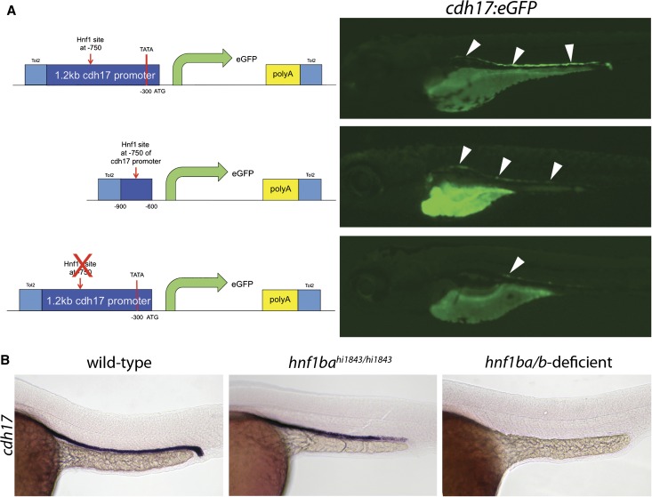
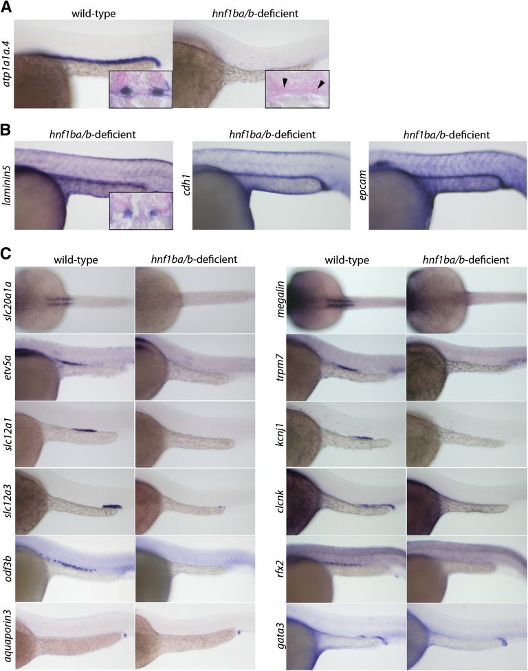
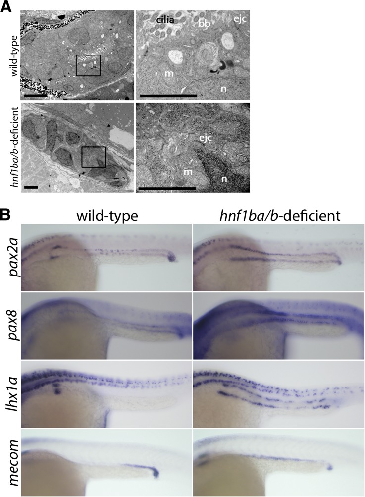
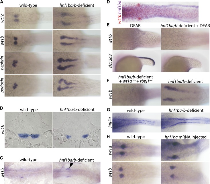
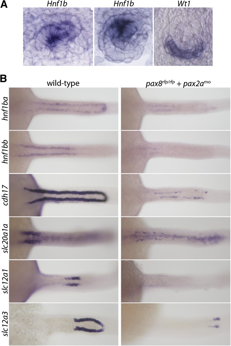
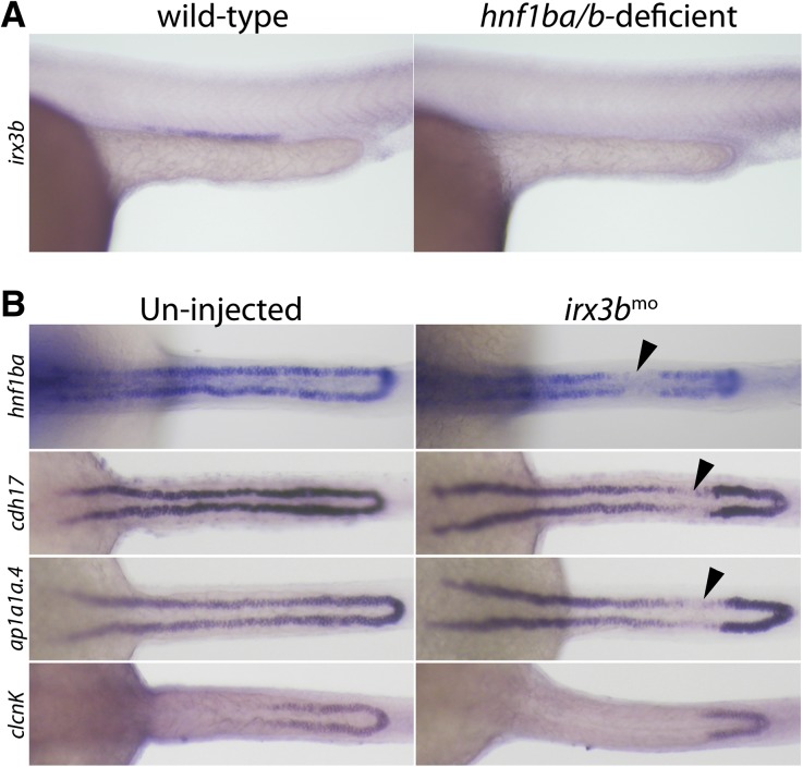
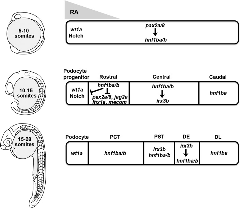
Nephrons comprise a blood filter and an epithelial tubule that is subdivided into proximal and distal segments, but what directs this patterning during kidney organogenesis is not well understood. Using zebrafish, we found that the HNF1β paralogues hnf1ba and hnf1bb, which encode homeodomain transcription factors, are essential for normal segmentation of nephrons. Embryos deficient in hnf1ba and hnf1bb did not express proximal and distal segment markers, yet still developed an epithelial tubule. Initiating hnf1ba/b expression required Pax2a and Pax8, but hnf1ba/b-deficient embryos did not exhibit the expected downregulation of pax2a and pax8 at later stages of development, suggesting complex regulatory loops involving these molecules. Embryos deficient in hnf1ba/b also did not express the irx3b transcription factor, which is responsible for differentiation of the first distal tubule segment. Reciprocally, embryos deficient in irx3b exhibited downregulation of hnf1ba/b transcripts in the distal early segment, suggesting a segment-specific regulatory circuit. Deficiency of hnf1ba/b also led to ectopic expansion of podocytes into the proximal tubule domain. Epistasis experiments showed that the formation of podocytes required wt1a, which encodes the Wilms' tumor suppressor-1 transcription factor, and rbpj, which encodes a mediator of canonical Notch signaling, downstream or parallel to hnf1ba/b. Taken together, these results suggest that Hnf1β factors are essential for normal segmentation of nephrons during kidney organogenesis.


Figures

Figure 1.
Figure 1.
Hnf1b regulates endogenous cdh17 expression. (A) 1.2 kb of genomic sequence upstream from the first exon of the cdh17 locus is able to drive eGFP expression in the pronephros of transiently transgenic zebrafish embryos (arrowheads). A 300 bp fragment of this cdh17 promoter sequence can recapitulate eGFP fluorescence observed in the full 1.2 kb promoter. Deletion of an Hnf1 binding motif at position −750 of the full 1.2 kb cdh17 promoter severely reduces transgenesis (embryos are photographed at 5 dpf). (B) At 24 hpf, cdh17 expression in hnf1bahi1843/hi1843 embryos is reduced in the distal segments of the pronephros compared with wild-type embryos. hnf1bbmo knockdown in hnf1bahi1843/hi1843 embryos (hnf1ba/b-deficient) completely inhibits cdh17 expression in the pronephros.
Figure 2.
Figure 2.
Nephron segmentation fails to occur in hnf1ba/b-deficient embryos. (A) atp1a1a.4 is expressed in all tubule segments of wild-type pronephroi at 24 hpf, but is not expressed in hnf1ba/b-deficient embryos. Sectioning of hnf1ba/b-deficient embryos in situ hybridized for atp1a1a.4 reveals the presence of a tubule (inset, arrowheads). (B) laminin5, cdh1, and epcam expression is normal in hnf1ba/b-deficient embryos, and laminin5 is clearly present in tubules of sectioned embryos (inset). (C) Hnf1ba/b-deficient animals lack expression of slc20a1a and megalin (PCT segment), etv5a and trpm7 (PST segment), slc12a1 and kcnj1 (DE segment), slc12a3 (DL segment), clcnk (DE and DL segments), and odf3b and rfx2 (markers of multi-cilliated cells). aquaporin3 (marker of the cloaca) expression is unaffected by hnf1ba/b-deficiency. gata3 expression in corpuscle of stannius cells is lost in hnf1ba/b-deficient embryos, but is maintained in the duct segment. (All photos are lateral views except for slc20a1a and megalin, which are dorsal views.)
Figure 3.
Figure 3.
Epithelial characteristics are retained, but early acting renal regulators are not downregulated in hnf1ba/b-deficient embryos. (A) Electron microscopy images of 3 dpf wild-type (top panels) and hnf1ba/b-deficient (bottom panels) embryos sectioned through the proximal tubule region. Black box indicates the area magnified in the right-hand panels. (B) Expression of early acting renal regulators pax2a, pax8, lhx1a, and mecom are maintained in all segments of the pronephros of hnf1ba/b-deficient embryos. bb, brush border; ejc, epithelial junctional complex; m, mitochondria; n, nucleus. Scale bars, 0.5 μm.
Figure 4.
Figure 4.
Ectopic podocyte formation occurs in hnf1ba/b-deficient embryos. (A) wt1a and wt1b are ectopically expressed in the proximal tubule domain of hnf1ba/b-deficient embryos at 24 hpf. Similarly, nephrin+ and podocin+ cells are expanded in hnf1ba/b -deficient embryos at 48 hpf. (B) Normal midline fusion of the wt1b+ podocytes at 24 hpf does not occur in hnf1ba/b-deficient embryos. (C) At the 15-somite stage of development, we observe an expansion of wt1b+ cells in hnf1ba/b -deficient embryos (arrowhead). (D) At the 15-somite stage, wt1b and hnf1ba expression are not overlapping and mark distinct regions of the renal epithelium (wt1b+ podocytes and hnf1ba+ tubules). (E) hnf1ba/b deficiency does not rescue loss of wt1b+ podocytes in DEAB-treated embryos. Expansion of DL tubule marker slc12a3 into proximal tubule segments of DEAB-treated embryos is observed, but in hnf1ba/b-deficient embryos treated with DEAB, slc12a3 expression is completely prevented. (F) Morpholino knockdown of wt1a and rbpj1 prevents formation of ectopic wt1b+ cells in hnf1ba/b-deficient embryos. (G) jagged2a (jag2a) expression is maintained in the proximal pronephros of hnf1ba/b deficient embryos. (H) hnf1ba mRNA overexpression does not suppress wt1a+ and wt1b+ cell formation.
Figure 5.
Figure 5.
Mouse Hnf1b is expressed in distal portions of S-shaped bodies and pax2a/pax8-doubly deficient embryos have reduced expression of nephron segment markers. (A) In E15.5 mice, Hnf1b is distally restricted in comma-shaped bodies (left panel) and Hnf1b and Wt1 show nonoverlapping expression domains in S-shaped bodies (right panels). (B) pax8rfp/rfp/pax2amo embryos show reduced expression of hnf1ba, hnf1bb, cdh17, slc20a1a, slc12a1, and slc12a3 at 24 hpf.
Figure 6.
Figure 6.
Irx3b regulates hnf1ba expression in the DE segment. (A) hnf1ba/b-deficient embryos do not express irx3b in the central region of the pronephric tubules. (B) irx3b-deficient embryos are created by morpholino knockdown. In these embryos, we observe reduced hnf1ba, cdh17, atp1a1a.4, and clcnk expression in the DE segment (arrowheads).
Figure 7.
Figure 7.
A model for the spatiotemporal regulation of nephron segmentation by Hnf1b factors in zebrafish.

Similar articles

Cited by

References

    1. Wingert RA, Davidson AJ: The zebrafish pronephros: A model to study nephron segmentation. Kidney Int 73: 1120–1127, 2008 - PubMed
    1. Wingert RA, Selleck R, Yu J, Song HD, Chen Z, Song A, Zhou Y, Thisse B, Thisse C, McMahon AP, Davidson AJ: The cdx genes and retinoic acid control the positioning and segmentation of the zebrafish pronephros. PLoS Genet 3: 1922–1938, 2007 - PMC - PubMed
    1. Dawid IB, Breen JJ, Toyama R: LIM domains: Multiple roles as adapters and functional modifiers in protein interactions. Trends Genet 14: 156–162, 1998 - PubMed
    1. Pfeffer PL, Gerster T, Lun K, Brand M, Busslinger M: Characterization of three novel members of the zebrafish Pax2/5/8 family: Dependency of Pax5 and Pax8 expression on the Pax2.1 (noi) function. Development 125: 3063–3074, 1998 - PubMed
    1. Toyama R, Kobayashi M, Tomita T, Dawid IB: Expression of LIM-domain binding protein (ldb) genes during zebrafish embryogenesis. Mech Dev 71: 197–200, 1998 - PubMed

Publication types

MeSH terms