. 2012 Dec;23(12):2001-11.
doi: 10.1681/ASN.2012020139. Epub 2012 Nov 15.

Deleting the TGF-β receptor attenuates acute proximal tubule injury

Affiliations

Deleting the TGF-β receptor attenuates acute proximal tubule injury

Leslie Gewin et al. J Am Soc Nephrol. 2012 Dec.

Abstract

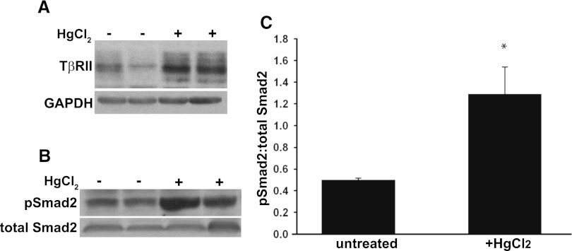
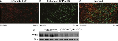
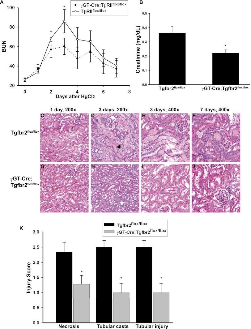
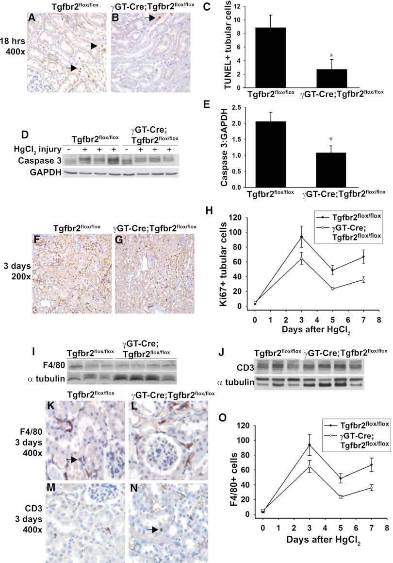
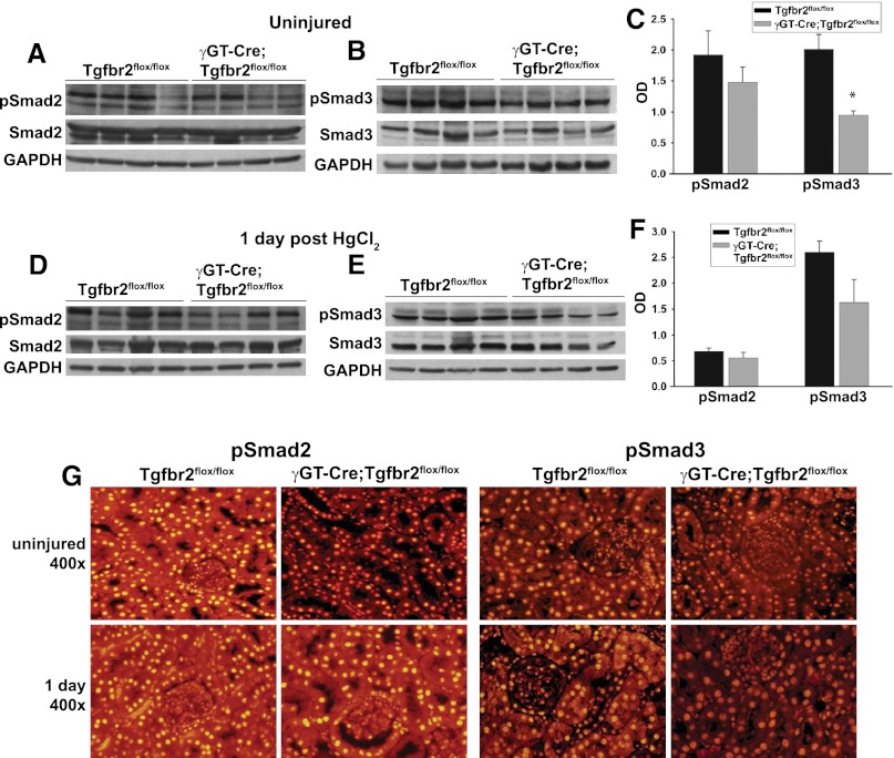
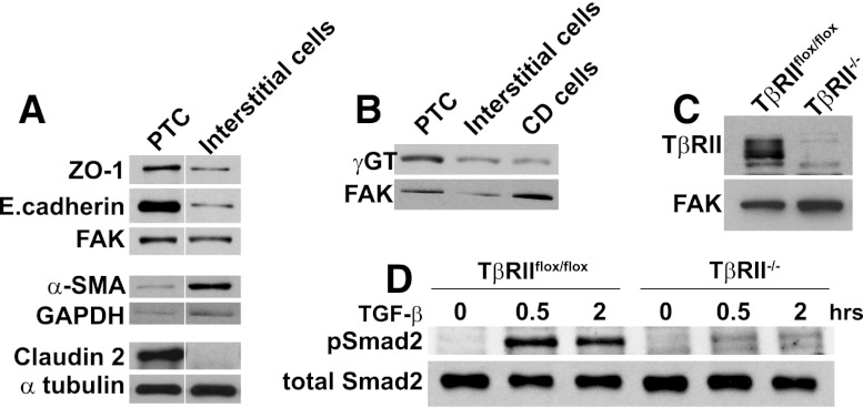
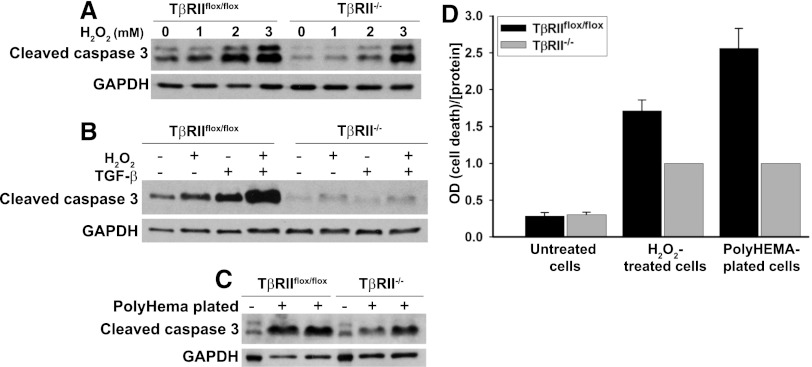
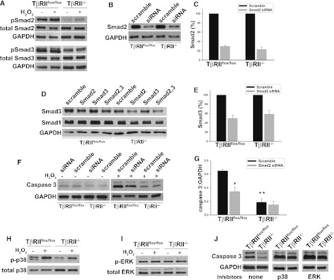
TGF-β is a profibrotic growth factor in CKD, but its role in modulating the kidney's response to AKI is not well understood. The proximal tubule epithelial cell, which is the main cellular target of AKI, expresses high levels of both TGF-β and its receptors. To determine how TGF-β signaling in this tubular segment affects the response to AKI, we selectively deleted the TGF-β type II receptor in the proximal tubules of mice. This deletion attenuated renal impairment and reduced tubular apoptosis in mercuric chloride-induced injury. In vitro, deficiency of the TGF-β type II receptor protected proximal tubule epithelial cells from hydrogen peroxide-induced apoptosis, which was mediated in part by Smad-dependent signaling. Taken together, these results suggest that TGF-β signaling in the proximal tubule has a detrimental effect on the response to AKI as a result of its proapoptotic effects.


Figures

Figure 1.
Figure 1.
HgCl2 stimulated TGF-β signaling in renal cortices. (A) Renal cortices were dissected from wild-type FVB mice that were injected with HgCl2 3 days earlier or were untreated. The tissue lysates were immunoblotted for TβRII expression. (B) Tissue lysates of renal cortices isolated 1 day after HgCl2 were immunoblotted for pSmad2 and total Smad2. (C) Bands from six mice (three treated, three untreated) were quantified by densitometry and reported as means ± SEMs. *P<0.05. GADPH, glyceraldehyde 3-phosphate dehydrogenase.
Figure 2.
Figure 2.
Deletion of TβRII in the γGT-Cre;Tgfbr2flox/flox mice. The mT/mG reporter mouse was crossed with our γGT-Cre;Tgfbr2flox/flox mice, demonstrating membrane-bound red fluorescence (mT) in all cells except those where Cre is active and green fluorescence (mG) replaces mT (A and B). (C) The red (580–610 nm) and green (510–540 nm) wavelengths are merged, showing that Cre is primarily expressed in tubules in the cortex and corticomedullary renal tissue. (D) Cortical lysates of adult Tgfbr2flox/flox and γGT-Cre;Tgfbr2flox/flox mice were immunoblotted for TβRII expression with focal adhesion kinase (FAK) used as loading control.
Figure 3.
Figure 3.
γGT-Cre;Tgfbr2flox/flox mice were protected from HgCl2-induced injury. (A) Plasma BUN levels (mg/dl) were measured at the time of injury and daily thereafter until euthanasia at day 7. Means are shown ± SEMs from 14 Tgfbr2flox/flox and 12 γGT-Cre;Tgfbr2flox/flox mice. *P<0.05. (B) Plasma creatinine 3 days after HgCl2 injection was measured by HPLC on six Tgfbr2flox/flox and five γGT-Cre;Tgfbr2flox/flox mice. Means are shown ± SEMs. *P<0.05. (C–J) Tissue from mice at 1, 3, and 7 days after treatment with HgCl2 was stained with hematoxylin and eosin. There was minimal epithelial injury 1 day after HgCl2, but at 3 days, there was significantly more cast formation (black arrow in D) and tubular necrosis in the Tgfbr2flox/flox than in the γGT-Cre;Tgfbr2flox/flox mice. Both genotypes had resolving epithelial injury at 7 days, but fewer residual tubular casts were present in the γGT-Cre;Tgfbr2flox/flox mice. (K) Injury was scored at 3 days, as discussed in the Concise Methods section. The values represent the means of six Tgfbr2flox/flox and seven γGT-Cre;Tgfbr2flox/flox mice. Means are shown ± SEMs. *P<0.05.
Figure 4.
Figure 4.
γGT-Cre;Tgfbr2flox/flox mice had less tubular apoptosis after HgCl2. (A and B) TUNEL staining was performed at 18 hours after injury with apoptotic nuclei staining brown (arrows). (C) TUNEL-positive tubular cells from 10 hpf per mouse were counted in a blinded fashion and reported as means using five mice per genotype ± SEMs. *P<0.05. (D) Cortical kidney lysates 18 hours after HgCl2 injections were immunoblotted for cleaved caspase 3 expression using glyceraldehyde 3-phosphate dehydrogenase (GAPDH) as a loading control. Three representative mice per genotype are shown. (E) Bands were quantified by densitometry expressed as cleaved caspase 3:GAPDH means from five Tgfbr2flox/flox and six γGT-Cre;Tgfbr2flox/flox mice ± SEMs. *P<0.05. (F–H) Ki-67 staining was performed on five mice per genotype at 0, 3, 5, and 7 days after HgCl2 treatment. (F and G) Representative staining at 3 days is shown. (H) Ki-67+ tubular cells were counted in 10 hpf per mouse at the different time points and expressed as means ± SEMs. (I and J) Cortical tissue lysates from three wild-type and four γGT-Cre;Tgfbr2flox/flox mice at 3 days after HgCl2 injection were immunoblotted for F4/80 and CD3 to detect macrophages and T cells, respectively. Representative staining of F4/80 (K and L) and CD3 (M and N) at 3 days is shown with positively staining cells in brown (arrows). F4/80 staining was performed on four mice per genotype at days 0, 3, and 7, and F4/80+ cells were counted in 10 hpf per mouse and shown as means ± SEMs (O).
Figure 5.
Figure 5.
Smad activity is suppressed in γGT-Cre;Tgfbr2flox/flox mice. (A–F) Cortical tissue lysates from wild-type and γGT-Cre;Tgfbr2flox/flox mice at 0 and 1 day after HgCl2 were immunoblotted for phosphorylated Smad2 (pSmad2), total Smad2, phosphorylated Smad3 (pSmad3), total Smad3, and glyceraldehyde 3-phosphate dehydrogenase (GAPDH) for loading. (C, F) Densitometry on phosphorylated Smads2/3 was normalized to GAPDH and reported as means ± SEMs. *P<0.05. (G) Localization of pSmad2/3 was determined by immunofluorescence on paraffin-embedded tissue at 0 and 1 day after injury.
Figure 6.
Figure 6.
PTCs were generated and TβRII deleted in vitro. (A and B) Cell lysates of PTCs, interstitial cells, and collecting duct (CD) cells were immunoblotted for expression of zona occludens-1 (ZO-1), E.cadherin, α-smooth muscle actin (α-SMA), claudin 2, and γ−glutamyltransferase (γGT) with focal adhesion kinase (FAK), glyceraldehyde 3-phosphate dehydrogenase (GAPDH), and α tubulin as loading controls. Vertical white lines separate samples rearranged from the same immunoblot. (C) Deletion of TβRII was confirmed by immunoblots of TβRIIflox/flox and TβRII−/− PTCs. (D) Cell lysates of TβRIIflox/flox and TβRII−/− PTC treated with TGF-β1 (2.5 ng/ml) for various time points were immunoblotted for phosphorylated and total levels of Smad2.
Figure 7.
Figure 7.
TβRII−/− PTC are resistant to apoptosis. (A) TβRIIflox/flox and TβRII−/− PTCs were treated with various concentrations of H2O2 for 12 hours, and then immunoblots for cleaved caspase 3 were performed with glyceraldehyde 3-phosphate dehydrogenase (GAPDH) as a loading control. (B) PTCs were treated with H2O2 (1mM) or TGF-β1 (2.5 ng/ml) for 12 hours and immunoblotted for cleaved caspase 3 and GAPDH. (C) TβRIIflox/flox and TβRII−/− PTCs were plated on PolyHEMA-coated plates, as described in the Concise Methods section, for 18 hours to induce anoikis and then immunoblotted for cleaved caspase 3 and GAPDH. (D) The Roche Cell Death ELISA was used to quantify apoptosis in PTCs both treated with H2O2 and plated on PolyHEMA. Experiments were repeated three times, and ratios of cell death to protein concentration were normalized to 1 for TβRII−/− PTCs. The values for untreated cells (negative controls) are expressed relative to TβRII−/− H2O2-treated PTCs as means ± SEMs.
Figure 8.
Figure 8.
Increased apoptosis in TβRIIflox/flox PTCs is partly Smad dependent. (A) Smad2/3 phosphorylation was measured in PTCs at basal conditions and after 30 minutes of H2O2 (1 mM). Vertical white lines separate samples rearranged from the same blot. (B and C) PTCs were transfected with 25 nM Smad2 or scramble siRNA as discussed in the Concise Methods, and knockdown of protein expression was confirmed 4 days later by immunoblots. (C) Smad2 protein expression was quantified by densitometry, normalized to loading controls, and expressed as a percentage of scramble siRNA-treated cells based on the average from three different experiments ± SEM. (D) Smad3 and Smad1 protein expression was measured after transfection with Smad2 (Ambion) with our without Smad3 (Dharmacon) siRNA. (E) Smad2 siRNA reduced expression of Smad3 as quantified by densitometry on three experiments, normalized to glyceraldehyde 3-phosphate dehydrogenase (GAPDH) and expressed as a percentage of scramble siRNA ± SEM. (F and G) Lysates of TβRIIflox/flox and TβRII−/− PTCs transfected with Smad or scramble siRNA and with or without H2O2 (1 mM) for 12 hours were immunoblotted for cleaved caspase 3 (Caspase 3) and GAPDH as a loading control. (G) Densitometry was performed on three different experiments with the average OD readings shown for H2O2-treated PTCs ± SEMs. *P<0.01 for apoptosis of scramble- versus Smad-siRNA–treated TβRIIflox/flox PTCs. **P<0.01 for the difference in apoptosis between TβRIIflox/flox and TβRII−/− PTCs after H2O2. (H and I) PTCs with or without 2 mM H2O2 were immunoblotted for total and phosphorylated p38 and for ERK. (J) TβRIIflox/flox and TβRII−/− PTCs treated with no inhibitors, 10 μM p38 inhibitor (SB203582), or 10 μM ERK inhibitor (UO126) were incubated with H2O2 (2 mM) for 12 hours and immunoblotted for cleaved caspase 3 (Caspase 3). The vertical white lines separate samples rearranged in order from the same immunoblot.

Similar articles

Cited by

References

    1. Humes HD, Cieslinski DA, Coimbra TM, Messana JM, Galvao C: Epidermal growth factor enhances renal tubule cell regeneration and repair and accelerates the recovery of renal function in postischemic acute renal failure. J Clin Invest 84: 1757–1761, 1989 - PMC - PubMed
    1. Coimbra TM, Cieslinski DA, Humes HD: Epidermal growth factor accelerates renal repair in mercuric chloride nephrotoxicity. Am J Physiol 259: F438–F443, 1990 - PubMed
    1. Miller SB, Martin DR, Kissane J, Hammerman MR: Insulin-like growth factor I accelerates recovery from ischemic acute tubular necrosis in the rat. Proc Natl Acad Sci U S A 89: 11876–11880, 1992 - PMC - PubMed
    1. Miller SB, Martin DR, Kissane J, Hammerman MR: Hepatocyte growth factor accelerates recovery from acute ischemic renal injury in rats. Am J Physiol 266: F129–F134, 1994 - PubMed
    1. Wrana JL, Attisano L, Cárcamo J, Zentella A, Doody J, Laiho M, Wang XF, Massagué J: TGF beta signals through a heteromeric protein kinase receptor complex. Cell 71: 1003–1014, 1992 - PubMed

Publication types

MeSH terms

Substances