. 2013 Mar;62(3):905-11.
doi: 10.2337/db12-0600. Epub 2012 Nov 16.

Local autoantigen expression as essential gatekeeper of memory T-cell recruitment to islet grafts in diabetic hosts

Affiliations

Local autoantigen expression as essential gatekeeper of memory T-cell recruitment to islet grafts in diabetic hosts

Gonnie M Alkemade et al. Diabetes. 2013 Mar.

Abstract

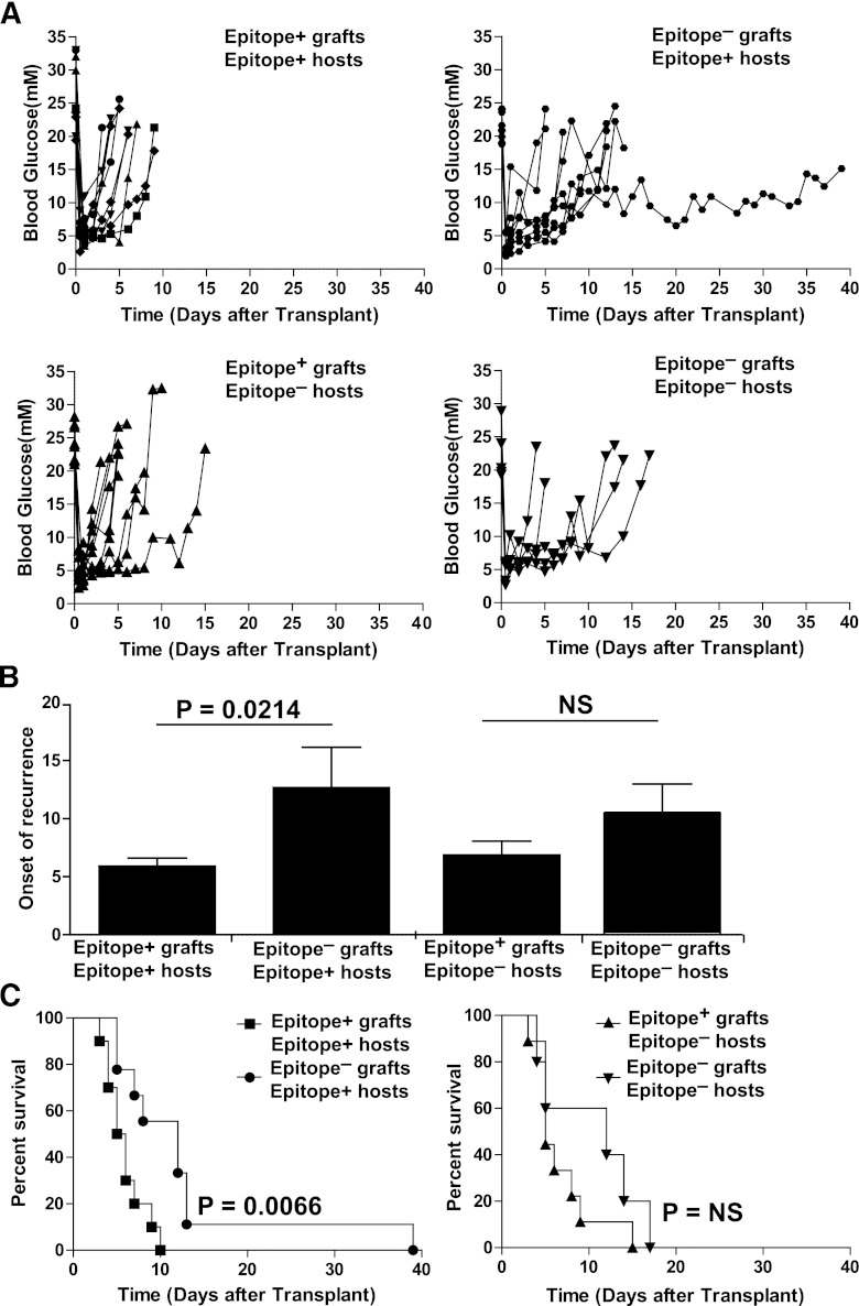
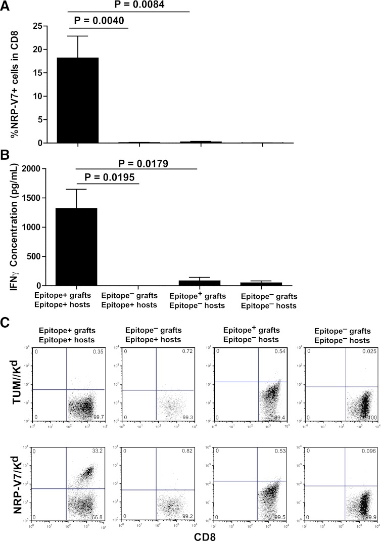
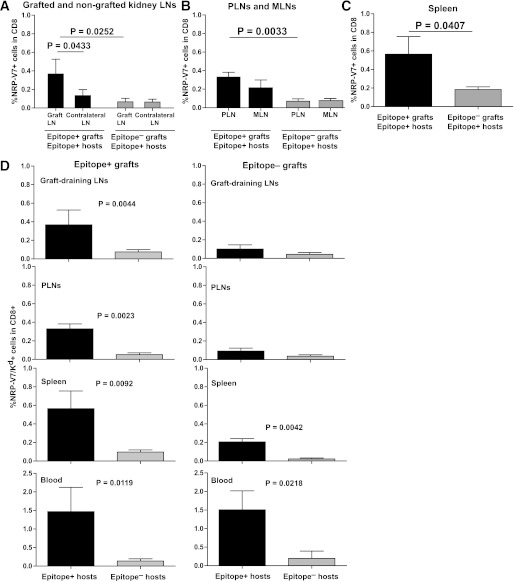
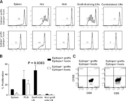
It is generally believed that inflammatory cues can attract noncognate, "bystander" T-cell specificities to sites of inflammation. We have shown that recruitment of naive and in vitro activated autoreactive CD8⁺ T cells into endogenous islets requires local autoantigen expression. Here, we demonstrate that absence of an autoantigen in syngeneic extrapancreatic islet grafts in diabetic hosts renders the grafts "invisible" to cognate memory (and naive) T cells. We monitored the recruitment of islet-specific glucose-6-phosphatase catalytic subunit-related protein (IGRP)₂₀₆₋₂₁₄-reactive CD8⁺ T cells into IGRP₂₀₆₋₂₁₄-competent and IGRP₂₀₆₋₂₁₄-deficient islet grafts in diabetic wild-type or IGRP₂₀₆₋₂₁₄(-/-) nonobese diabetic hosts (harboring either naive and memory T cells or only naive IGRP₂₀₆₋₂₁₄-specific T-cells, respectively). All four host-donor combinations had development of recurrent diabetes within 2 weeks. Wild-type hosts recruited IGRP₂₀₆₋₂₁₄-specific T cells into IGRP₂₀₆₋₂₁₄(+/+) but not IGRP₂₀₆₋₂₁₄(-/-) grafts. In IGRP₂₀₆₋₂₁₄(-/-) hosts, there was no recruitment of IGRP₂₀₆₋₂₁₄-specific T cells, regardless of donor type. Graft-derived IGRP₂₀₆₋₂₁₄ activated naive IGRP₂₀₆₋₂₁₄-specific T cells, but graft destruction invariably predated their recruitment. These results indicate that recurrent diabetes is exclusively driven by autoreactive T cells primed during the primary autoimmune response, and demonstrate that local antigen expression is a sine qua non requirement for accumulation of memory T cells into islet grafts. These findings underscore the importance of tackling autoreactive T-cell memory after β-cell replacement therapy.


Figures

FIG. 1.
FIG. 1.
Survival of islet grafts from IGRP206–214-competent or IGRP206–214-deficient donors in spontaneously diabetic IGRP206–214-competent or IGRP206–214-deficient NOD hosts. A: Individual blood glucose curves of diabetic NOD hosts receiving NOD.scid (n = 10) or NOD.rag2KI/KI.IGRPK209A/F213AKI/KI islets (n = 9), and diabetic NOD.IGRPK209A/F213AKI/KI hosts receiving NOD.scid (n = 9) or NOD.rag2−/−.IGRPK209A/F213AKI/KI islets (n = 5). B: Average onset of disease recurrence after transplantation (in days) in the four different donor/host combinations. P values were obtained by Mann-Whitney U test. C: Survival curves of grafts in diabetic NOD (left) or NOD.IGRPK209A/F213AKI/KI hosts (right). P values were calculated via log-rank test. For (B) and (C), differences between epitope-positive and epitope-negative grafts in epitope-positive hosts remained statistically significant upon exclusion of the epitope-negative graft that survived to 40 days (P = 0.0395 in B; and P = 0.0353 in C).
FIG. 2.
FIG. 2.
Recruitment of IGRP206–214-reactive CD8+ T cells from diabetic IGRP206–214-competent or IGRP206–214-deficient NOD hosts into islet grafts from IGRP206–214-competent or IGRP206–214-deficient donors. A: Percentages of NRP-V7/Kd tetramer-positive in islet-graft–associated CD8+ T cells. Data (average ± SEM) correspond, from left to right, to eight, four, four, and three grafts per group, respectively. B: Interferon-γ (IFN-γ) secretion by islet graft-associated CD8+ T cells in response to NRP-V7 peptide-pulsed NOD dendritic cells. Data correspond, from left to right, to five, four, four, and three grafts per group, respectively. C: Representative fluorescence-activated cell sorting staining profiles of CD8+ T cells isolated from islet grafts in the four different donor/host combinations. TUM/Kd was used as a negative control tetramer. P values in (A) and (B) were obtained with Mann-Whitney U test. Grafts were harvested immediately after the last blood glucose measurement in Fig. 1. (A high-quality color representation of this figure is available in the online issue.)
FIG. 3.
FIG. 3.
Frequencies of IGRP206–214-reactive CD8+ T cells in lymphoid organs and blood of diabetic IGRP206–214-competent or IGRP206–214-deficient hosts grafted with IGRP206–214-competent or IGRP206–214-deficient islets. Percentages of NRP-V7/Kd tetramer-positive cells in CD8+ T cells from lymph nodes (LNs) draining the grafted compared with contralateral kidneys (A), the pancreatic lymph nodes (PLNs) and the mesenteric lymph nodes (MLNs) (B), and the spleen (C). Data (average ± SEM) correspond to six and eight mice, respectively. D: Frequencies of IGRP206–214-reactive CD8+ T cells in lymphoid organs and blood of IGRP206–214-competent compared with IGRP206–214-deficient hosts grafted with IGRP206–214-competent (left) compared with IGRP206–214-deficient islets (right). Data (average ± SEM) correspond to six, six, eight, and five mice per group, respectively. Background staining with the negative control tetramer TUM/Kd was subtracted. P values were obtained with Mann-Whitney U test.
FIG. 4.
FIG. 4.
Proliferation of naive CFSE-labeled 8.3-CD8+ T cells in lymphoid organs of IGRP206–214-competent hosts grafted with IGRP206–214-competent compared with IGRP206–214-deficient islets. A: Representative CFSE dilution profiles. B: Average ± SEM of the percentage of proliferated cells. C: Representative flow profiles of graft-associated CD8+ T cells of these mice. Data in (AC) correspond to three mice per host/donor combination. P values were obtained with Mann-Whitney U test. LN, lymph node; MLN, mesenteric lymph node; PLN, pancreatic lymph node.

References

    1. Babad J, Geliebter A, DiLorenzo TP. T-cell autoantigens in the non-obese diabetic mouse model of autoimmune diabetes. Immunology 2010;131:459–465 - PMC - PubMed
    1. Anderson B, Park BJ, Verdaguer J, Amrani A, Santamaria P. Prevalent CD8(+) T cell response against one peptide/MHC complex in autoimmune diabetes. Proc Natl Acad Sci USA 1999;96:9311–9316 - PMC - PubMed
    1. DiLorenzo T, Graser T, Ono T, et al. Major histocompatibility complex class I-restricted T cells are required for all but end stages of diabetes development in nonobese diabetic mice and use a prevalent T cell receptor alpha chain gene rearrangement. Proc Natl Acad Sci USA 1998;95:12538–12543 - PMC - PubMed
    1. Verdaguer J, Yoon JW, Anderson B, et al. Acceleration of spontaneous diabetes in TCR-beta-transgenic nonobese diabetic mice by beta-cell cytotoxic CD8+ T cells expressing identical endogenous TCR-alpha chains. J Immunol 1996;157:4726–4735 - PubMed
    1. Verdaguer J, Schmidt D, Amrani A, Anderson B, Averill N, Santamaria P. Spontaneous autoimmune diabetes in monoclonal T cell nonobese diabetic mice. J Exp Med 1997;186:1663–1676 - PMC - PubMed

Publication types

MeSH terms