

Flow cytometry enables a high-throughput homogeneous fluorescent antibody-binding assay for cytotoxic T cell lytic granule exocytosis
- PMID: 23160568
- PMCID: PMC4043149
- DOI: 10.1177/1087057112466697
Flow cytometry enables a high-throughput homogeneous fluorescent antibody-binding assay for cytotoxic T cell lytic granule exocytosis
Abstract
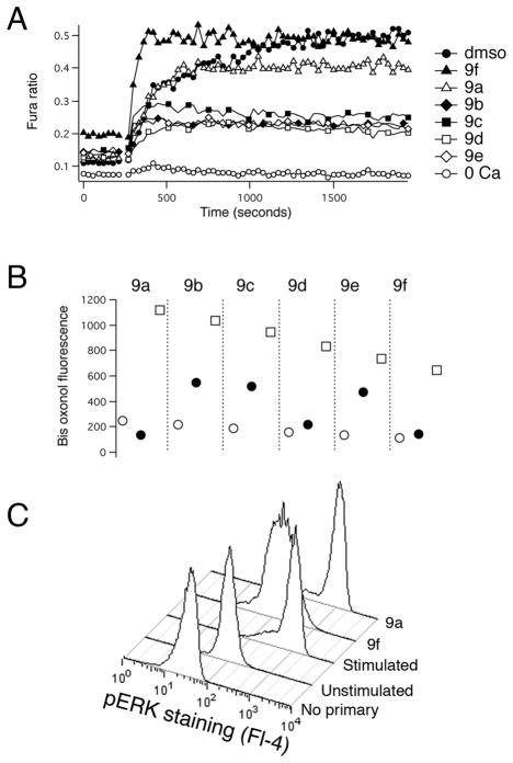
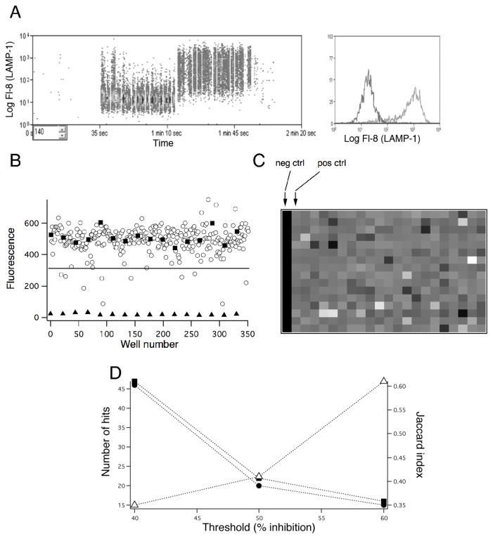
We developed a homogeneous phenotypic fluorescence end-point assay for cytotoxic T lymphocyte lytic granule exocytosis. This flow cytometric assay measures binding of an antibody to a luminal epitope of a lysosomal membrane protein (LAMP-1) that is exposed by exocytosis to the extracellular solution. Washing to remove unbound antibody is not required. Confirming the assay's ability to detect novel active compounds, we screened at a concentration of 50 µM a synthetic diversity library of 91 compounds in a 96-well plate format, identifying 17 compounds that blocked by 90% or more. The actions of six structurally related tetracyano-hexahydroisoindole compounds that inhibited by ~90% at a concentration of 10 µM were investigated further. Four reduced elevations in intracellular Ca(2+); it is likely that depolarization of the cells' membrane potential underlies the effect for at least two of the compounds. Another compound was found to be a potent inhibitor of the activation of the mitogen-activated protein (MAP) kinase ERK. Finally, we transferred the assay to a 384-well format and screened the Prestwick Compound Library using high-throughput flow cytometry. Our results indicate that our assay will likely be a useful means of screening libraries for novel compounds with important biological activities.
Figures


Similar articles
-
A high-throughput phenotypic screen of cytotoxic T lymphocyte lytic granule exocytosis reveals candidate immunosuppressants.J Biomol Screen. 2015 Mar;20(3):359-71. doi: 10.1177/1087057114557620. Epub 2014 Nov 7. J Biomol Screen. 2015. PMID: 25381253 Free PMC article.
-
Development of an Enhanced Phenotypic Screen of Cytotoxic T-Lymphocyte Lytic Granule Exocytosis Suitable for Use with Synthetic Compound and Natural Product Collections.J Biomol Screen. 2016 Jul;21(6):556-66. doi: 10.1177/1087057116643260. Epub 2016 Apr 5. J Biomol Screen. 2016. PMID: 27048485
-
A phenotypic screen for compounds that reverse cAMP-mediated suppression of T cell functions.SLAS Discov. 2022 Jul;27(5):314-322. doi: 10.1016/j.slasd.2022.03.008. Epub 2022 Apr 3. SLAS Discov. 2022. PMID: 35385793
-
Qualitative and quantitative analysis of subpopulations of cytotoxic effector cells by flow cytometry.J Lipid Mediat Cell Signal. 1994 Feb;9(1):19-25. J Lipid Mediat Cell Signal. 1994. PMID: 8032712 Review.
-
Computational analysis of high-throughput flow cytometry data.Expert Opin Drug Discov. 2012 Aug;7(8):679-93. doi: 10.1517/17460441.2012.693475. Epub 2012 Jun 18. Expert Opin Drug Discov. 2012. PMID: 22708834 Free PMC article. Review.
Cited by
-
Discussion Addendum for: Phosphine-Catalyzed [4 + 2] Annulation: Synthesis of Ethyl 6-Phenyl-1-tosyl-1,2,5,6-tetrahydropyridine-3-carboxylate.Organic Synth. 2019;96:110-123. Epub 2019 Apr 22. Organic Synth. 2019. PMID: 31736515 Free PMC article.
-
Flow Cytometry: Impact on Early Drug Discovery.J Biomol Screen. 2015 Jul;20(6):689-707. doi: 10.1177/1087057115578273. Epub 2015 Mar 24. J Biomol Screen. 2015. PMID: 25805180 Free PMC article. Review.
-
Phosphine Organocatalysis.Chem Rev. 2018 Oct 24;118(20):10049-10293. doi: 10.1021/acs.chemrev.8b00081. Epub 2018 Sep 27. Chem Rev. 2018. PMID: 30260217 Free PMC article. Review.
-
Development of a screening strategy for new modulators of T cell receptor signaling and T cell activation.Sci Rep. 2018 Jul 3;8(1):10046. doi: 10.1038/s41598-018-28106-5. Sci Rep. 2018. PMID: 29968737 Free PMC article.
-
Advances in nucleophilic phosphine catalysis of alkenes, allenes, alkynes, and MBHADs.Chem Commun (Camb). 2013 Dec 25;49(99):11588-619. doi: 10.1039/c3cc47368f. Chem Commun (Camb). 2013. PMID: 24196409 Free PMC article.
References
-
- Johnston PA, Johnston PA. Cellular platforms for HTS: three case studies. Drug Discov Today. 2002;7:353–363. - PubMed
-
- An WF, Tolliday N. Cell-based assays for high-throughput screening. Mol Biotechnol. 2010;45:180–186. - PubMed
-
- Giepmans BN, Adams SR, Ellisman MH, Tsien RY. The fluorescent toolbox for assessing protein location and function. Science. 2006;312:217–224. - PubMed
-
- Sklar LA, Finney DA. Analysis of ligand-receptor interactions with the fluorescence activated cell sorter. Cytometry. 1982;3:161–165. - PubMed
Publication types
MeSH terms
Substances
Grants and funding
LinkOut - more resources
Full Text Sources
Miscellaneous
