Clinical Trial
. 2013 Jan;54(1):78-82.
doi: 10.2967/jnumed.112.111922. Epub 2012 Nov 15.

Imaging changes in synaptic acetylcholine availability in living human subjects

Affiliations
Clinical Trial

Imaging changes in synaptic acetylcholine availability in living human subjects

Irina Esterlis et al. J Nucl Med. 2013 Jan.

Abstract

In vivo estimation of β(2)-nicotinic acetylcholine receptor availability with molecular neuroimaging is complicated by competition between the endogenous neurotransmitter acetylcholine and the radioligand (123)I-3-[2(S)-2-azetidinylmethoxy]pyridine ((123)I-5-IA). We examined whether binding of (123)I-5-IA is sensitive to increases in extracellular levels of acetylcholine in humans, as suggested in nonhuman primates.

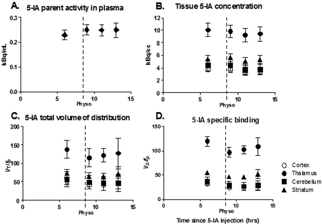
Methods: Six healthy subjects (31 ± 4 y) participated in a (123)I-5-IA SPECT study. After baseline scans, physostigmine (1-1.5 mg) was administered intravenously over 60 min, and 9 additional scans were obtained.

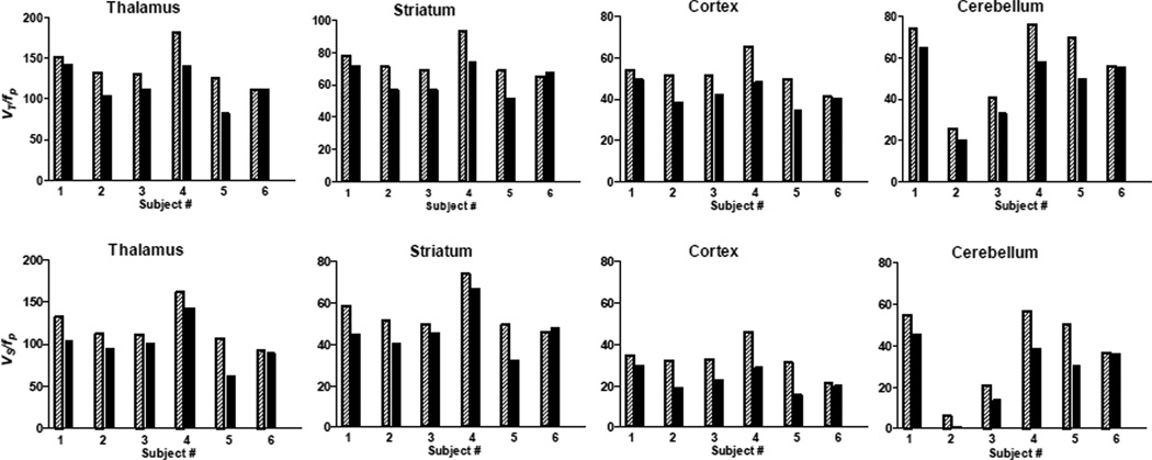
Results: We observed a significant reduction in the total volume of distribution after physostigmine administration (29% ± 17% in the cortex, 19% ± 15% in the thalamus, 19% ± 15% in the striatum, and 36% ± 30% in the cerebellum; P < 0.05). This reduction reflected a combination of a region-specific 7%-16% decrease in tissue concentration of tracer and a 9% increase in plasma parent concentration.

Conclusion: These data suggest that increases in acetylcholine compete with (123)I-5-IA for binding to β(2)-nicotinic acetylcholine receptor. Additional validation of this paradigm is warranted, but it may be used to interrogate changes in extracellular acetylcholine.


Figures

Figure 1
Figure 1
The first point in each graph represents baseline data obtained starting 6 h after beginning tracer infusion when a state of equilibrium is achieved, and provided the baseline specific binding. Following completion of the baseline scans, physostigmine was administered I.V. (1.0–1.5 mg over 1 h, arrow). At the onset of the physostigmine infusion, scanning was resumed for up to 9 h. Bars represent standard error of the mean (SEM). A. Plasma [123I]5-IA concentration (kBq/mL) (total parent) measured during [123I]5-IA constant infusion in healthy volunteers. Following physostigmine administration there was a significant 8% increase in mean plasma [123I]5-IA concentration as compared to before physostigmine administration. B. Tissue [123I]5-IA concentration (kBq/cc) in thalamus (circles), striatum (triangles), cortex (open circles), and cerebellum (squares) measured during [123I]5-IA constant infusion. We observed 7–15% region specific decrease in [123I]5-IA tissue concentration after physostigmine challenge. C. [123I]5-IA total volume of distribution (VT/fp) in thalamus (circles), striatum (triangles), cortex (open circles), and cerebellum (squares) measured during [123I]5-IA constant infusion. VT/fp values measured after the physostigmine infusion were significantly reduced (14–18% region specific) compared to the baseline values. D. [123I]5-IA specific binding (BPf) in thalamus (circles), striatum (triangles), cortex (open circles), and cerebellum (squares) measured during [123I]5-IA constant infusion. BPf values measured after the physostigmine infusion were significantly reduced (19–36% region specific) compared to the baseline values.
Figure 2
Figure 2
A. β2-nAChR availability (VT/fP) before (shaded bars) and after (solid bars) physostigmine injection for each subject. Thalamus: Percent displacement of 5-IA for subjects 1–6 were −7%, −22%, −14%, −22%, −35%, −0%, respectively. Striatum: Percent displacement of 5-IA for subjects 1–6 was −8%, −20%, −18%, −20%, −25%, −4%, respectively. Cortex: Percent displacement of 5-IA for subjects 1–6 were −9%, −25%, −18%, −26%, −30%, −2%, respectively. Cerebellum: Percent displacement of 5-IA for subjects 1–6 was −12%, −20%, −18%, −23%, −28%, 0%, respectively. B. Specific radioligand binding (Vs/fP) before (shaded bars) and after (solid bars) physostigmine injection for each subject. Thalamus: Percent displacement of 5-IA for subjects 1–6 were −8%, −25%, −17%, −25%, −41%, 0%, respectively. Striatum: Percent displacement of 5-IA for subjects 1–6 was −10%, −27%, −24%, −26%, −34%, +5%, respectively. Cortex: Percent displacement of 5-IA for subjects 1–6 were −13%, −40%, −29%, −36%, −50%, −6%, respectively. Cerebellum: Percent displacement of 5-IA for subjects 1–6 was −17%, −90%, − 35%, −32%, −40%, −1%, respectively.

Similar articles

Cited by

References

    1. Fujita M, Al-Tikriti M, Tamagnan G, et al. Influence of acetylcholine levels on the binding of a SPECT nicotinic acetylcholine receptor ligand [123I]5-I-A-85380. Synapse. 2003;48:116–122. - PubMed
    1. Saricicek A, Esterlis I, Maloney K, et al. Persistent β2*-Nicotinic Acetylcholinergic Receptor Dysfunction in Major Depressive Disorder. American Journal of Psychiatry. 2012 - PMC - PubMed
    1. Abreo M, Lin N-H, Garvey D, et al. Novel 3-Pyridyl Ethers with Subnanomolar Affinity for Central Neuronal Nicotinic Acetylcholine Receptors. J Med Chem. 1996;39:817–825. - PubMed
    1. Vaupel D, Mukhin A, Kimes A, Horti A, Koren A, London E. In vivo studies with [125I]5-IA 85380, a nicotinic acetylcholine receptor radioligand. NeuroReport. 1998;9:2311–2317. - PubMed
    1. Mukhin A, Gundisch D, Horti A, Koren A, Tamagnan G, Kimes A, et al. 5-Iodo-A-85830, an α4β2 subtype-selective ligand for nicotinic acetylcholine receptors. Mol Pharmacol. 2000;57:642–649. - PubMed

Publication types

LinkOut - more resources