. 2013 Jan;11(1):86-94.
doi: 10.1158/1541-7786.MCR-12-0243-T. Epub 2012 Nov 16.

Regulation of CXCR4-mediated invasion by DARPP-32 in gastric cancer cells

Affiliations

Regulation of CXCR4-mediated invasion by DARPP-32 in gastric cancer cells

Shoumin Zhu et al. Mol Cancer Res. 2013 Jan.

Abstract

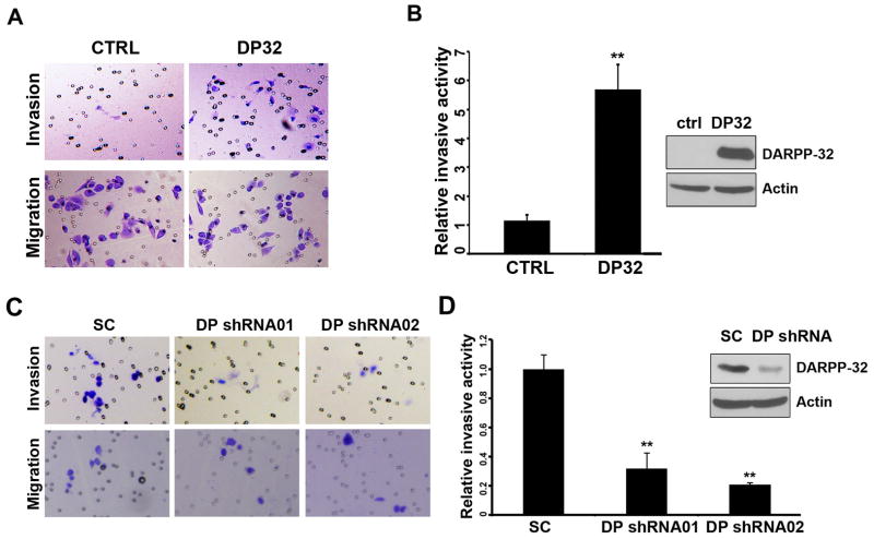
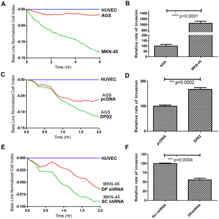
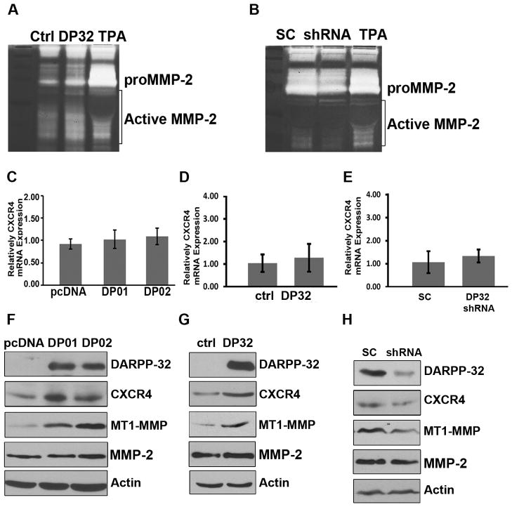
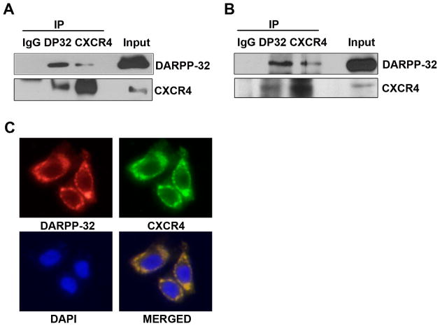
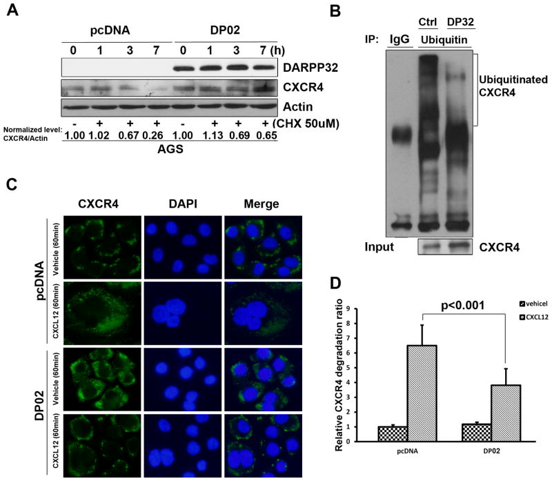
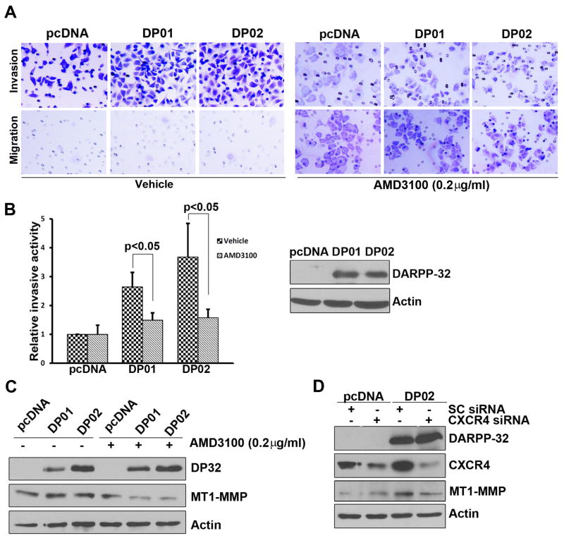
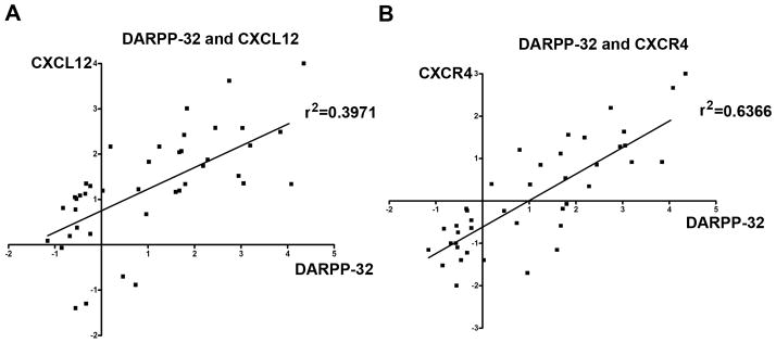
Although Dopamine and cAMP-regulated phosphoprotein, Mr 32000 (DARPP-32) is overexpressed in two-thirds of gastric cancers, its impact on molecular functions has not been fully characterized. In this study, we examined the role of DARPP-32 in gastric cancer cell invasion. Using matrigel-coated Boyden chamber invasion assay, DARPP-32-overexpressing AGS cells showed a three-fold increase in invasion relative to the vector control (P < 0.01). We also tested the transendothelial cell invasion as a measure of cell aggressiveness using the impedance-based human umbilical vein endothelial cells invasion assay and obtained similar results (P < 0.001). Western blot analysis indicated that overexpression of DARPP-32 mediated an increase in the membrane-type 1 matrix metalloproteinase (MT1-MMP) and CXCR4 protein levels. Consistent with the role of MT1-MMP in cleaving extracellular matrix proteins initiating the activation of soluble MMPs, we detected a robust increase in MMP-2 activity in DARPP-32-overexpressing cells. The knockdown of endogenous DARPP-32 in the MKN-45 cells reversed these signaling events and decreased cell invasive activity. We tested whether the invasive activity mediated by DARPP-32 might involve sustained signaling via CXCR4-dependent activation of the MT1-MMP/MMP-2 pathway. The small-molecule CXCR4 antagonist (AMD3100) and CXCR4-siRNA blocked DARPP-32-induced cell invasion. We further examined our hypothesis that DARPP-32 could interact with CXCR4 and stabilize its levels following stimulation with its ligand, CXCL12. Using reciprocal coimmunoprecipitation and immunofluorescence experiments, we found that DARPP-32 and CXCR4 coexist in the same protein complex. DARPP-32 prolonged the CXCR4 protein half-life and reduced ubiquitination of the CXCR4 protein, following treatment with its ligand, CXCL12. In conclusion, these findings show a novel mechanism by which DARPP-32 promotes cell invasion by regulating CXCR4-mediated activation of the MT1-MMP/MMP-2 pathway.


Conflict of interest statement

Conflict of interest

The authors declare no conflict of interest.

Figures

Figure 1
Figure 1. DARPP-32 increased invasive activity in gastric cancer cells
A) Invasion assays were performed using the matrigel-boyden chamber assay. AGS cells lentiviral expressing DARPP-32 (DP32) are more invasive than control (ctrl). B) The quantification demonstrates a significant increase in the number of invasive cells in AGS cells stably expressing DARPP-32 (p<0.01). Western blot demonstrates the levels of DARPP-32. C–D) The knockdown of DARPP-32 in the MKN-45 cells (shRNA01 and 02) led to a significant reduction in cell invasion (p<0.01). The levels of DARPP-32 are shown by Western blot analysis.
Figure 2
Figure 2. DARPP-32 enhances gastric cancer cells invasion
A–B) 5000 cells each of AGS or MKN-45 were used to challenge the HUVEC monolayer. The cell index was measured every 5 min by the xCELLigence system. The rate of change of cell index as a function of time was calculated as a measure of invasive activity. MKN45 cells have a higher change in cell index than AGS cells. C–D) The same as in A–B, using AGS cells stably expressing DARPP-32 or pcDNA demonstrates a significant increase in the rate of change of cell index in DARPP-32 expressing AGS cells (DP02) as compared to control (pcDNA) (p=0.0002). E–F) The same as in A–B, following knockdown of DARPP-32 (DP shRNA) or control (SC shRNA) in MKN45 cells, demonstrates a significant decrease in the rate of change of cell index (P=0.0004).
Figure 3
Figure 3. DARPP-32 enhances the chemokine receptor CXCR4 expression
A) Gelatin zymography of AGS cells expressing lentiviral particles of DARPP-32 (DP32) demonstrates activation of MMP2 as compared to control particles (ctrl), TPA was used as a positive control. B) The lentiviral shRNA knockdown of endogenous DARPP-32 in MKN45 cells reversed these findings. The qRT-PCR analysis demonstrates non-significant differences in the level of CXCR4 in AGS cells stably overexpressing pcDNA-DARPP-32 (DP01 and DP02) (C) or lentivirus particles of DARPP-32 (D), as compared to controls. E) Similarly, no differences were observed following knockdown of endogenous DARPP-32 in MKN45 cells. Western blot analysis of AGS cells stably expressing DARPP-32 (F) or lentiviral DARPP-32particles (G) demonstrates up-regulation of CXCR4 and MT1-MMP, but not MMP-2. H) The lentiviral shRNA knockdown of endogenous DARPP-32 in MKN45 cells reversed these findings.
Figure 4
Figure 4. DARPP-32 interacts with CXCR4
A) HEK-293 cells were transiently transfected with DARPP-32 and CXCR4. Two-way co-immunoprecipitation and Western blot analyses demonstrates the interaction of DARPP-32 and CXCR4. B) Similar to (A), the protein interaction of endogenous DARPP-32 and CXCR4 was evaluated by co-immunoprecipitation in MKN-45 cells. C) Immunofluorescence analysis using AGS cells, following overexpression of DARPP-32, demonstrates co-localization of CXCR4 and DARPP-32 proteins on the cell membrane.
Figure 5
Figure 5. DARPP-32 promotes CXCR4 protein stability
A) Western blot analysis, following the treatment with CHX (50 uM) for the indicated time points, demonstrated increased levels of CXCR4 protein in AGS cells stably expressing DARPP-32, as compared to control (pcDNA). B) HEK-293 cells co-transfected with HA-CXCR4, His-ubiquitin and pcDNA or DARPP-32 were treated with 30 nM CXCL-12 for 30 min. Cell lysates were incubated with an anti-HA polyclonal antibody and the immunoprecipitates were analyzed by SDS-PAGE followed by Western blotting using an anti-His antibody conjugated to HRP. Cell lysates were analyzed for the presence of HA-CXCR4 and demonstrated less CXCR4 ubiquitination in DARPP-32-expressing cells, as compared to control. C) AGS cells stably expressing DARPP-32 (DP02) or empty vector were treated with 30 nM CXCL-12 or vehicle for 60 min, followed by immunofluorescence staining with anti-CXCR4 (green) and DAPI (blue). CXCR4 remained mainly localized to the plasma membrane after vehicle or CXCL-12 treatment of DARPP-32-expressing AGS cells. On the contrary, CXCR4 was localized to diffuse punctate cytosolic vesicles in control cells D) Quantification of relative CXCR4 degradation shows a significant decrease in the relative CXCR4 degradation in the AGS cells stably-expressing DARPP-32 (p<0.001).
Figure 6
Figure 6. Blocking of CXCR4 disrupts DARPP-32-mediated cancer cells invasion
A) Invasive assays were performed in the AGS cells-stably expressing DARPP-32 after treatment with AMD3100 (0.2 μg/ml) or vehicle. B) Quantification of invasion assay shows a significant decrease in relative cell invasion after AMD3100 treatment (p<0.05), the levels of DARPP-32 are shown in the Western blots. C) Western blot analysis following the above conditions demonstrates a decrease in the levels of MT1-MMP in DARPP-32 expressing cells (DP01 and 02). D) The knockdown of CXCR4 (CXCR4 siRNA) in AGS cells stably expressing DARPP-32 led to a decrease in the levels of CXCR4 and MT1-MMP, as compared to control (Sc siRNA).
Figure 7
Figure 7. Positive correlation between expression levels of DARPP-32, CXCL-12 and CXCR-4
The qRT-PCR analysis of the expression levels of CXCL-12, CXCR4, and DARPP-32 in gastric tumors and their adjacent histologically normal non-tumor tissue samples demonstrated a statistically significant positive correlation between the levels of CXCL-12 and DARPP-32 (A) and between CXCR4 and DARPP-32 (B), suggesting that these components of invasion activity co-exist in gastric cancer cells.

Similar articles

Cited by

References

    1. Ferlay J, Shin HR, Bray F, Forman D, Mathers C, Parkin DM. Estimates of worldwide burden of cancer in 2008: GLOBOCAN 2008. International journal of cancer Journal international du cancer. 2010;127:2893–917. - PubMed
    1. Catalano V, Labianca R, Beretta GD, Gatta G, de Braud F, Van Cutsem E. Gastric cancer. Crit Rev Oncol Hematol. 2009;71:127–64. - PubMed
    1. Nairn AC, Hemmings HC, Jr, Walaas SI, Greengard P. DARPP-32 and phosphatase inhibitor-1, two structurally related inhibitors of protein phosphatase-1, are both present in striatonigral neurons. J Neurochem. 1988;50:257–62. - PubMed
    1. Mukherjee K, Peng D, Brifkani Z, Belkhiri A, Pera M, Koyama T, et al. Dopamine and cAMP regulated phosphoprotein MW 32 kDa is overexpressed in early stages of gastric tumorigenesis. Surgery. 2010;148:354–63. - PMC - PubMed
    1. El-Rifai W, Smith MF, Jr, Li G, Beckler A, Carl VS, Montgomery E, et al. Gastric cancers overexpress DARPP-32 and a novel isoform, t-DARPP. Cancer Res. 2002;62:4061–4. - PubMed

Publication types

MeSH terms

Substances