. 2013 Feb;83(2):389-98.
doi: 10.1124/mol.112.081315. Epub 2012 Nov 15.

Functional characterization of three mouse formyl peptide receptors

Affiliations

Functional characterization of three mouse formyl peptide receptors

Hui-Qiong He et al. Mol Pharmacol. 2013 Feb.

Abstract

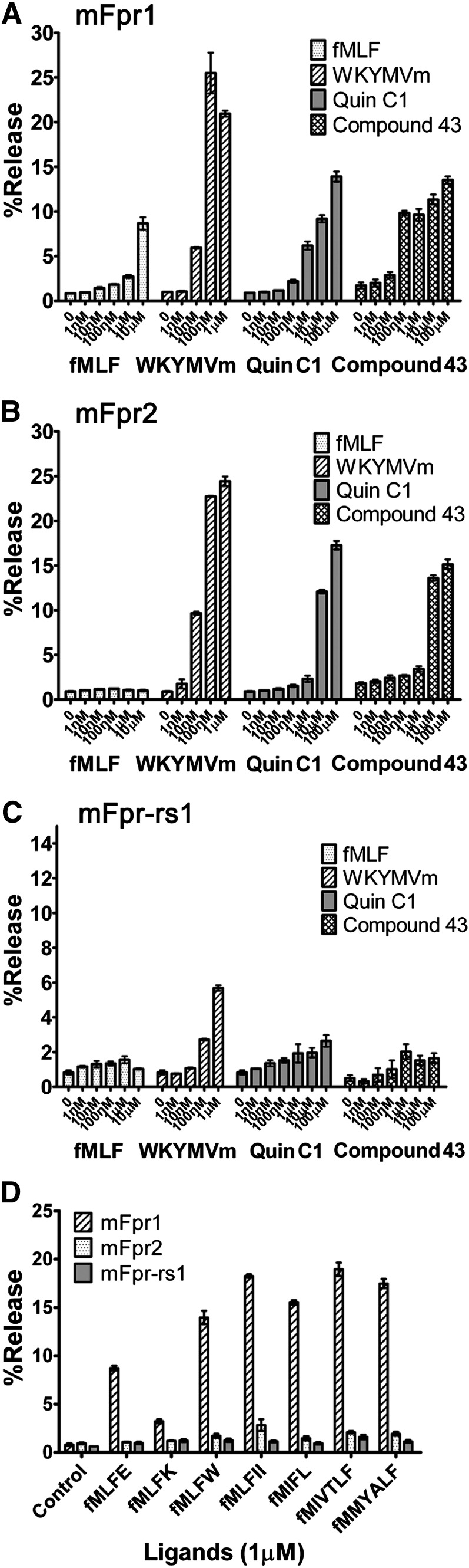
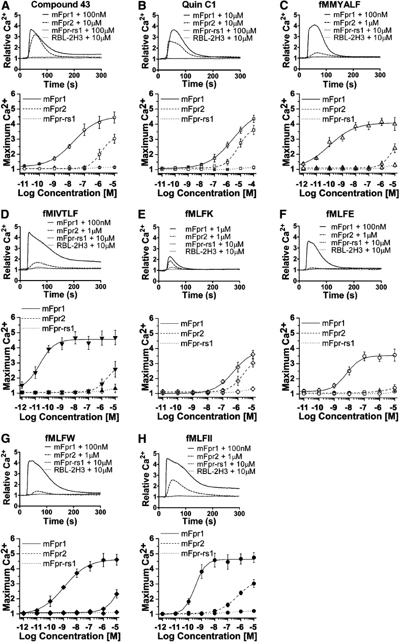
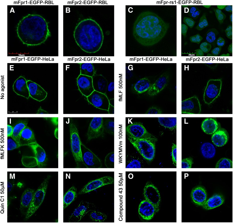
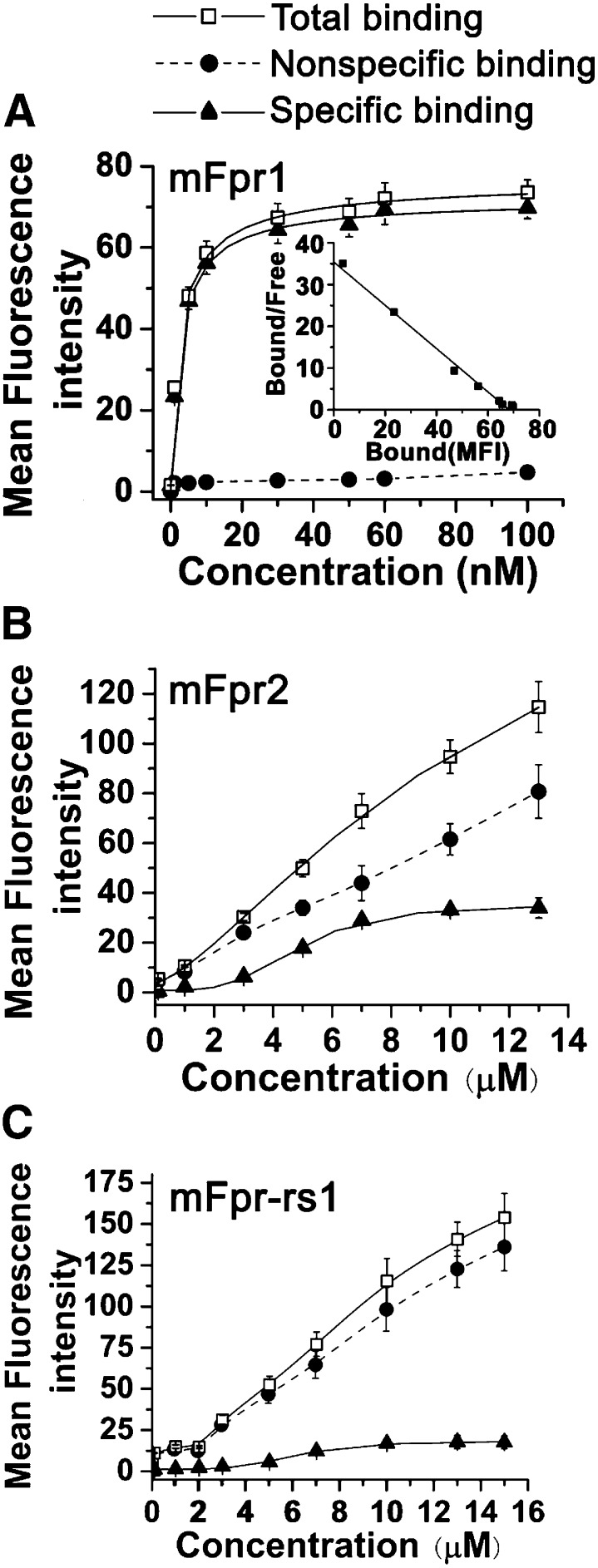
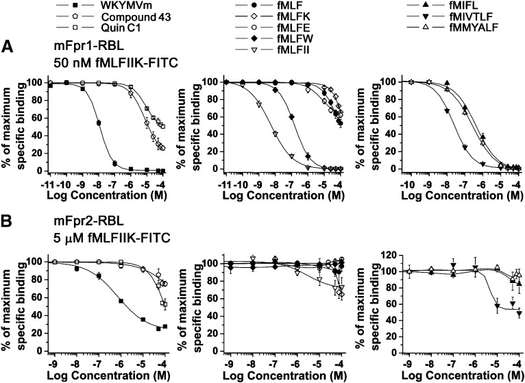
The evolutionary relationship and functional correlation between human formyl peptide receptors (FPRs) and their mouse counterparts remain incompletely understood. We examined three members of the mouse formyl peptide receptor subfamily (mFprs) and found that they differ in agonist preference and cellular distributions. When stably expressed in transfected rat basophilic leukemia (RBL-2H3) cells, mFpr1 was readily activated by N-formylated peptides derived from Listeria monocytogenes (fMIVTLF), Staphylococcus aureus (fMIFL), and mitochondria (fMMYALF). In contrast, the Escherichia coli-derived fMLF was 1000-fold less potent. The aforementioned peptides were much less efficacious at mFpr2, which responded better to the synthetic hexapeptide WKYMVm, the synthetic agonists Quin-C1 (a substituted quinazolinone), and compound 43 (a nitrosylated pyrazolone derivative). Saturation binding assays showed that mFpr1 and mFpr2 were expressed at similar levels on the cell surface, although their affinity for N-formyl-Met-Leu-Phe-Ile-Ile-Lys-fluorescein isothiocyanate varied by more than 1000-fold [dissociation constant (K(d)) values of 2.8 nM for mFpr1 and 4.8 μM for mFpr2]). Contrary to these receptors, mFpr-rs1 responded poorly to all the previously mentioned peptides that were tested. Fluorescent microscopy revealed an intracellular distribution pattern of mFpr-rs1. On the basis of these results, we conclude that mFpr1 is an ortholog of human FPR1 with certain pharmacologic properties of human FPR2/ALX, whereas mFpr2 has much lower affinity for formyl peptides. The intracellular distribution of mFpr-rs1 suggests an evolutionary correlation with human FPR3.


Figures

Fig. 1.
Fig. 1.
Degranulation induced by agonists through mFpr1, mFpr2 or mFpr-rs1 receptors. Release of β-hexosaminidase by fMLF, WKYMVm, Quin-C1, and compound 43 at indicated concentrations were measured in RBL cells expressing mFpr1 (A), mFpr2 (B), or mFpr-rs1 (C), respectively. Various formyl peptides (1 μM) were compared for the induction of β-hexosaminidase secretion (D). Values are mean ± S.E.M. of single duplicate determinations and representative of at least three separate experiments.
Fig. 2.
Fig. 2.
Calcium mobilization in RBL-mFpr cells stimulated with various agonists. Cells were loaded with FLIPR Calcium 5 reagent and analyzed for changes of intracellular calcium in response to agonist stimulation. (A) Quin-C1, (B) compound 43, (C) fMMYALF, (D) fMIVTLF, (E) fMLFK, (F) fMLFE, (G) fMLFW, and (H) fMLFII. The upper panels show typical calcium traces in response to the indicated agonist concentration. The lower panels are dose-dependent curves, which were based on peak Ca2+ increase at indicated agonist concentrations and shown as mean ± S.E.M. representing >3 separate experiments.
Fig. 3.
Fig. 3.
Cell surface expression and internalization of mouse Fpr receptors. (A) mFpr1-EGFP, (B) mFpr2-EGFP, or (C and D) mFpr-rs1-EGFP expressed in RBL cells. The scale bar for (A–C) is shown (A), and the images were captured using a 63× oil immersion objective, whereas (D) was captured under a 40× dry objective, and the scale bar is shown in the figure. Internalization of mFpr1-EGFP or mFpr2-EGFP in HeLa cells, before (E and F) and after 30 minutes of stimulation with fMLF (G and H), fMLFK (I and J), WKYMVm (K and L), Quin-C1 (M and N), or compound 43 (O and P), at indicated concentrations. The scale bar for (E–P) is shown in (E), and the images were captured using a 63× oil immersion objective.
Fig. 4.
Fig. 4.
Binding assays with RBL-2H3 cells stably transfected by mFpr1, mFpr2, or mFpr-rs1. Saturation, nonspecific binding, and specific binding of fMLFIIK-FITC to (A) mFpr1-, (B) mFpr2-, or (C) mFpr-rs1- expressing RBL cells. The inlet shows Scatchard analysis of the data in (A). Data were analyzed with Origin 7.5 software (Northampton, MA), and the results are shown as means ± S.E.M. with >3 experiments.
Fig. 5.
Fig. 5.
Competition binding assays performed on (A) mFpr1-RBL and (B) mFpr2-RBL cells. Binding of fMLFIIK-FITC (50 nM for mFpr1-RBL and 5 μM for mFpr2-RBL cells) was competitively displaced by increasing concentrations of agonists, including synthetic ligands WKYMVm, Quin-C1, and Compound 43 (left); E. coli-derived fMLF and derivatives fMLFK, fMLFE, fMLFW, and fMLFII (middle); and other bacterial formyl peptides fMIFL, fMIVTLF, and mitochondrial fMMYALF (right). Data were analyzed as described in the legend for Fig. 4. The results are shown as means ± S.E.M. with >3 experiments.
Fig. 6.
Fig. 6.
Comparison of formyl and nonformyl peptides in competitive binding assay. The ability of nonformylated peptide MLFIIK and N-formylated fMLFIIK to compete with fMLFIIK-FITC for binding to (A) mFpr1- and (B) mFpr2-RBL cells was determined. All data were analyzed as described in the legend of Fig. 4.

Similar articles

Cited by

References

    1. Bürli RW, Xu H, Zou X, et al. (2006) Potent hFPRL1 (ALXR) agonists as potential anti-inflammatory agents. Bioorg Med Chem Lett 16:3713–3718 - PubMed
    1. Chen K, Le Y, Liu Y, Gong W, Ying G, Huang J, Yoshimura T, Tessarollo L, Wang JM. (2010) A critical role for the g protein-coupled receptor mFPR2 in airway inflammation and immune responses. J Immunol 184:3331–3335 - PMC - PubMed
    1. Chiang N, Serhan CN, Dahlén SE, Drazen JM, Hay DW, Rovati GE, Shimizu T, Yokomizo T, Brink C. (2006) The lipoxin receptor ALX: potent ligand-specific and stereoselective actions in vivo. Pharmacol Rev 58:463–487 - PubMed
    1. Dufton N, Hannon R, Brancaleone V, Dalli J, Patel HB, Gray M, D’Acquisto F, Buckingham JC, Perretti M, Flower RJ. (2010) Anti-inflammatory role of the murine formyl-peptide receptor 2: ligand-specific effects on leukocyte responses and experimental inflammation. J Immunol 184:2611–2619 - PMC - PubMed
    1. Forsman H, Onnheim K, Andreasson E, Dahlgren C. (2011) What formyl peptide receptors, if any, are triggered by compound 43 and lipoxin A4? Scand J Immunol 74:227–234 - PubMed

Publication types

Substances

LinkOut - more resources