Review
. 2013 Aug;70(16):2899-917.
doi: 10.1007/s00018-012-1197-9. Epub 2012 Nov 16.

Co-ordinating Notch, BMP, and TGF-β signaling during heart valve development

Affiliations
Review

Co-ordinating Notch, BMP, and TGF-β signaling during heart valve development

Victoria C Garside et al. Cell Mol Life Sci. 2013 Aug.

Abstract

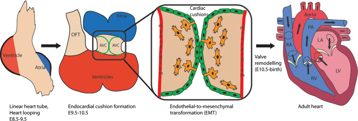
Congenital heart defects affect approximately 1-5 % of human newborns each year, and of these cardiac defects 20-30 % are due to heart valve abnormalities. Recent literature indicates that the key factors and pathways that regulate valve development are also implicated in congenital heart defects and valve disease. Currently, there are limited options for treatment of valve disease, and therefore having a better understanding of valve development can contribute critical insight into congenital valve defects and disease. There are three major signaling pathways required for early specification and initiation of endothelial-to-mesenchymal transformation (EMT) in the cardiac cushions: BMP, TGF-β, and Notch signaling. BMPs secreted from the myocardium set up the environment for the overlying endocardium to become activated; Notch signaling initiates EMT; and both BMP and TGF-β signaling synergize with Notch to promote the transition of endothelia to mesenchyme and the mesenchymal cell invasiveness. Together, these three essential signaling pathways help form the cardiac cushions and populate them with mesenchyme and, consequently, set off the cascade of events required to develop mature heart valves. Furthermore, integration and cross-talk between these pathways generate highly stratified and delicate valve leaflets and septa of the heart. Here, we discuss BMP, TGF-β, and Notch signaling pathways during mouse cardiac cushion formation and how they together produce a coordinated EMT response in the developing mouse valves.


Figures

Fig. 1
Fig. 1
Early development of the mouse embryonic heart. Initially, the heart forms a linear tube that generates swellings that create the future chambers of the heart at E8.5. At E9.0 the linear heart tube will begin looping to bring the chambers into their final positions. Endocardial cushion formation begins at E9.5 as AVC endocardial cells undergo endothelial-to-mesenchymal transformation (EMT) to create the AVC mesenchyme cells that fill the cardiac cushions and form the primitive heart valves. Following EMT (E10.5-birth), the primitive heart valves will remodel to generate the thin, delicate heart valve leaflets in the adult heart. In the first two panels, red depicts ventricles, and blue represents atria. The third panel is an inset from the E10.5 heart illustrating the AVC where red is the myocardium, green cells are endocardial cells, and orange cells are mesenchyme. The last panel depicts a cross section through the adult heart where red/pink represents oxygen-rich blood and blue represents oxygen-depleted blood. AVC atrioventricular canal, RA right atria, LA left atria, RV right ventricle, LV left ventricle, PA pulmonary artery
Fig. 2
Fig. 2
TGF-β, BMP, and Notch signaling pathways. a TGF-β and BMP signaling pathways. To activate signaling, a TGF-β or a BMP ligand binds their respective receptor complexes that contain two type I receptors and two type II receptors, which causes a phosphorylation event that activates the receptor Smads (R-Smads). Following this, R-Smads bind with their co-Smad, Smad4, and move into the nucleus to activate TGF-β or BMP-responsive genes, respectively. TGF-β Smads Smad2, Smad3. BMP Smads Smad1, Smad5, Smad8 (S1/5/8). The yellow star with a P represents a phosphorylation event. b Notch signaling pathway. Notch signaling is activated when a signaling cell with the Notch ligands, Jagged (Jag) or Delta-like (Dll) bind with a Notch receptor on a signal-receiving cell. The binding of the ligand to the Notch receptor causes a conformational change and exposes two cleavage sites S2 and S3 in the Notch receptor. The first cleavage of Notch occurs at the S2 site via a disintegrin and metalloproteinase (ADAM) protein that releases the extracellular portion of Notch. The second cleavage of Notch occurs at the S3 site by γ-secretase complex and this releases the intracellular domain of Notch (NICD). NICD translocates into the nucleus and binds with RBPJ (Recombination signal-binding protein 1 for J-Kappa), MAML (Master-mind like), p300, and co-activators (CoA) to activate Notch-responsive genes. In the absence of NICD, RBPJ is bound by co-repressors (CoR) and cannot activate Notch-responsive genes
Fig. 3
Fig. 3
Cross-talk between BMP, TGF-β, and Notch signaling pathways. Red represents Notch pathway-induced interactions. Blue represents BMP-induced interactions, and green represents TGF-β-induced interactions

Similar articles

Cited by

References

    1. Hoffman JI, Kaplan S. The incidence of congenital heart disease. J Am Coll Cardiol. 2002;39(12):1890–1900. doi: 10.1016/S0735-1097(02)01886-7. - DOI - PubMed
    1. Pierpont ME, Basson CT, Benson DW, Jr, Gelb BD, Giglia TM, Goldmuntz E, McGee G, Sable CA, Srivastava D, Webb CL. Genetic basis for congenital heart defects: current knowledge: a scientific statement from the American Heart Association Congenital Cardiac Defects Committee, Council on Cardiovascular Disease in the Young: endorsed by the American Academy of Pediatrics. Circulation. 2007;115(23):3015–3038. doi: 10.1161/CIRCULATIONAHA.106.183056. - DOI - PubMed
    1. Roger VL, Go AS, Lloyd-Jones DM, Adams RJ, Berry JD, Brown TM, Carnethon MR, Dai S, de Simone G, Ford ES, Fox CS, Fullerton HJ, Gillespie C, Greenlund KJ, Hailpern SM, Heit JA, Ho PM, Howard VJ, Kissela BM, Kittner SJ, Lackland DT, Lichtman JH, Lisabeth LD, Makuc DM, Marcus GM, Marelli A, Matchar DB, McDermott MM, Meigs JB, Moy CS, Mozaffarian D, Mussolino ME, Nichol G, Paynter NP, Rosamond WD, Sorlie PD, Stafford RS, Turan TN, Turner MB, Wong ND, Wylie-Rosett J. Heart disease and stroke statistics–2011 update: a report from the American Heart Association. Circulation. 2011;123(4):e18–e209. doi: 10.1161/CIR.0b013e3182009701. - DOI - PMC - PubMed
    1. Hinton RB, Jr, Lincoln J, Deutsch GH, Osinska H, Manning PB, Benson DW, Yutzey KE. Extracellular matrix remodeling and organization in developing and diseased aortic valves. Circ Res. 2006;98(11):1431–1438. doi: 10.1161/01.RES.0000224114.65109.4e. - DOI - PubMed
    1. Rabkin E, Aikawa M, Stone JR, Fukumoto Y, Libby P, Schoen FJ. Activated interstitial myofibroblasts express catabolic enzymes and mediate matrix remodeling in myxomatous heart valves. Circulation. 2001;104(21):2525–2532. doi: 10.1161/hc4601.099489. - DOI - PubMed

Publication types

MeSH terms

Substances

LinkOut - more resources