

CD133 Expression Strongly Correlates with the Phenotype of Very Small Embryonic-/Epiblast-Like Stem Cells
- PMID: 23161080
- PMCID: PMC5565199
- DOI: 10.1007/978-1-4614-5894-4_9
CD133 Expression Strongly Correlates with the Phenotype of Very Small Embryonic-/Epiblast-Like Stem Cells
Abstract
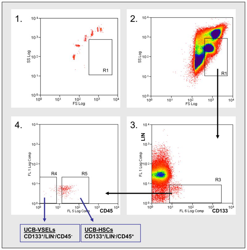
CD133 antigen (prominin-1) is a useful cell surface marker of very small embryonic-like stem cells (VSELs). Antibodies against it, conjugated to paramagnetic beads or fluorochromes, are thus powerful biological tools for their isolation from human umbilical cord blood, mobilized peripheral blood, and bone marrow. VSELs are described with the following characteristics: (1) are slightly smaller than red blood cells; (2) display a distinct morphology, typified by a high nuclear/cytoplasmic ratio and an unorganized euchromatin; (3) become mobilized during stress situations into peripheral blood; (4) are enriched in the CD133(+)Lin(-)CD45(-) cell fraction in humans; and (5) express markers of pluripotent stem cells (e.g., Oct-4, Nanog, and stage-specific embryonic antigen-4). The most recent in vivo data from our and other laboratories demonstrated that human VSELs exhibit some characteristics of long-term repopulating hematopoietic stem cells and are at the top of the hierarchy in the mesenchymal lineage. However, still more labor is needed to characterize better at a molecular level these rare cells.
Figures


Similar articles
-
Molecular and phenotypic characterization of CD133 and SSEA4 enriched very small embryonic-like stem cells in human cord blood.Leukemia. 2015 Sep;29(9):1909-17. doi: 10.1038/leu.2015.100. Epub 2015 Apr 17. Leukemia. 2015. PMID: 25882698
-
Very small embryonic/epiblast-like stem cells (VSELs) and their potential role in aging and organ rejuvenation--an update and comparison to other primitive small stem cells isolated from adult tissues.Aging (Albany NY). 2012 Apr;4(4):235-46. doi: 10.18632/aging.100449. Aging (Albany NY). 2012. PMID: 22498452 Free PMC article. Review.
-
Hematopoietic differentiation of umbilical cord blood-derived very small embryonic/epiblast-like stem cells.Leukemia. 2011 Aug;25(8):1278-85. doi: 10.1038/leu.2011.73. Epub 2011 Apr 12. Leukemia. 2011. PMID: 21483440 Free PMC article.
-
Umbilical cord blood-derived very small embryonic like stem cells (VSELs) as a source of pluripotent stem cells for regenerative medicine.Pediatr Endocrinol Rev. 2012 Mar;9(3):639-43. Pediatr Endocrinol Rev. 2012. PMID: 22523831 Review.
-
Very small embryonic-like stem-cell optimization of isolation protocols: an update of molecular signatures and a review of current in vivo applications.Exp Mol Med. 2013 Nov 15;45(11):e56. doi: 10.1038/emm.2013.117. Exp Mol Med. 2013. PMID: 24232255 Free PMC article. Review.
Cited by
-
Similar Population of CD133+ and DDX4+ VSEL-Like Stem Cells Sorted from Human Embryonic Stem Cell, Ovarian, and Ovarian Cancer Ascites Cell Cultures: The Real Embryonic Stem Cells?Cells. 2019 Jul 11;8(7):706. doi: 10.3390/cells8070706. Cells. 2019. PMID: 31336813 Free PMC article.
-
The proper criteria for identification and sorting of very small embryonic-like stem cells, and some nomenclature issues.Stem Cells Dev. 2014 Apr 1;23(7):702-13. doi: 10.1089/scd.2013.0472. Epub 2014 Jan 11. Stem Cells Dev. 2014. PMID: 24299281 Free PMC article. Review.
-
A Simplified and Effective Approach for the Isolation of Small Pluripotent Stem Cells Derived from Human Peripheral Blood.Biomedicines. 2023 Mar 5;11(3):787. doi: 10.3390/biomedicines11030787. Biomedicines. 2023. PMID: 36979766 Free PMC article.
-
HIF-2α and Oct4 have synergistic effects on survival and myocardial repair of very small embryonic-like mesenchymal stem cells in infarcted hearts.Cell Death Dis. 2017 Jan 12;8(1):e2548. doi: 10.1038/cddis.2016.480. Cell Death Dis. 2017. PMID: 28079892 Free PMC article.
-
Molecular and phenotypic characterization of CD133 and SSEA4 enriched very small embryonic-like stem cells in human cord blood.Leukemia. 2015 Sep;29(9):1909-17. doi: 10.1038/leu.2015.100. Epub 2015 Apr 17. Leukemia. 2015. PMID: 25882698
References
-
- D'Ippolito G, Diabira S, Howard GA, Menei P, Roos BA, Schiller PC. Marrow-isolated adult multilineage inducible (MIAMI) cells, a unique population of postnatal young and old human cells with extensive expansion and differentiation potential. Journal of Cell Science. 2004;117:2971–81. - PubMed
-
- Pittenger MF, Mackay AM, Beck SC, Jaiswal RK, Douglas R, Mosca JD, et al. Multilineage potential of adult human mesenchymal stem cells. Science. 1999;284:143–7. - PubMed
-
- Beltrami AP, Cesselli D, Bergamin N, Marcon P, Rigo S, Puppato E, et al. Multipotent cells can be generated in vitro from several adult human organs (heart, liver, and bone marrow) Blood. 2007;110:3438–46. - PubMed
-
- Vacanti MP, Roy A, Cortiella J, Bonassar L, Vacanti CA. Identification and initial characterization of spore-like cells in adult mammals. Journal of Cellular Biochemistry. 2001;80:455–60. - PubMed
MeSH terms
Grants and funding
LinkOut - more resources
Full Text Sources
Other Literature Sources
Research Materials
Miscellaneous
