Minimally invasive plate osteosynthesis of distal tibial fracture using a posterolateral approach: a cadaveric study and preliminary report
- PMID: 23161109
- PMCID: PMC3532637
- DOI: 10.1007/s00264-012-1712-5
Minimally invasive plate osteosynthesis of distal tibial fracture using a posterolateral approach: a cadaveric study and preliminary report
Abstract
Purpose: The aims of this anatomical study were to evaluate the feasibility of minimally invasive plate osteosynthesis (MIPO) using a posterolateral approach in distal tibial fractures and to study the relationship between neurovascular structures and the plate.
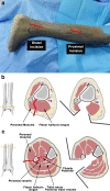
Methods: Two separate incisions, one proximal and one distal, were made on the posterolateral aspect of ten cadaveric legs in the prone position. A 14-hole contralateral anterolateral distal tibial locking plate was inserted into the submuscular tunnel using a posterolateral approach, and one screw was fixed on each side of the proximal and distal tibia. The MIPO tunnel was then explored to identify the relationship between neurovascular bundles and plate.
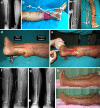
Results: For the proximal incision, retraction of the flexor hallucis longus and the tibialis posterior muscles medially was very important because it could protect the posterior tibial artery and the tibial nerve during plating. The sural nerve and lesser saphenous vein were easily identified and retracted in the superficial layer of the distal incision. In addition, we achieved satisfactory outcomes after using this MIPO technique in one patient.
Conclusion: Based on the results of our study, it seems that using the MIPO technique through a posterolateral approach should be a reasonable and safe treatment option for distal tibial fractures, especially when the anterior soft tissue is compromised. However, studies with a higher level of evidence should be done in more patients to confirm the clinical safety of using this technique.
Figures


Comment in
-
Minimally invasive plate osteosynthesis of distal tibial fracture using a posterolateral approach: a cadaveric study and preliminary report.Int Orthop. 2013 May;37(5):989-90. doi: 10.1007/s00264-013-1838-0. Epub 2013 Mar 7. Int Orthop. 2013. PMID: 23467894 Free PMC article. No abstract available.
-
Reply to comment on Kritsaneephaiboon et al. "Minimally invasive plate osteosynthesis of distal tibial fracture using a posterolateral approach: a cadaveric study and preliminary report".Int Orthop. 2013 May;37(5):991. doi: 10.1007/s00264-013-1840-6. Epub 2013 Mar 7. Int Orthop. 2013. PMID: 23467895 Free PMC article. No abstract available.
-
Reply to the comment on Kritsaneephaiboon et al.: Minimally invasive plate osteosynthesis of distal tibial fracture using a posterolateral approach: a cadaveric study and preliminary report.Int Orthop. 2013 Jun;37(6):1203-4. doi: 10.1007/s00264-013-1899-0. Epub 2013 May 1. Int Orthop. 2013. PMID: 23636836 Free PMC article. No abstract available.
-
Comment on Kritsaneephaiboon et al.: Minimally invasive plate osteosynthesis of distal tibial fracture using a posterolateral approach: a cadaveric study and preliminary report.Int Orthop. 2013 Jul;37(7):1423. doi: 10.1007/s00264-013-1918-1. Epub 2013 May 12. Int Orthop. 2013. PMID: 23665656 Free PMC article. No abstract available.
Similar articles
-
Clinical and radiological outcome of Gustilo type III open distal tibial and tibial shaft fractures after staged treatment with posterolateral minimally invasive plate osteosynthesis (MIPO) technique.Arch Orthop Trauma Surg. 2018 Aug;138(8):1097-1102. doi: 10.1007/s00402-018-2950-9. Epub 2018 May 10. Arch Orthop Trauma Surg. 2018. PMID: 29748878
-
The safety and feasibility of minimally invasive plate osteosynthesis (MIPO) on the medial side of the femur: A cadaveric injection study.Injury. 2015 Nov;46(11):2170-6. doi: 10.1016/j.injury.2015.08.032. Epub 2015 Aug 31. Injury. 2015. PMID: 26343301
-
Minimally invasive posteromedial percutaneous plate osteosynthesis for diaphyseal tibial fractures: technique description.Injury. 2017 Oct;48 Suppl 4:S6-S9. doi: 10.1016/S0020-1383(17)30768-4. Injury. 2017. PMID: 29145970
-
Minimally Invasive Plate Osteosynthesis for Distal Tibia Fractures.Clin Podiatr Med Surg. 2018 Apr;35(2):223-232. doi: 10.1016/j.cpm.2017.12.005. Epub 2018 Feb 1. Clin Podiatr Med Surg. 2018. PMID: 29482791 Review.
-
Intramedullary Nailing Versus Minimally Invasive Plate Osteosynthesis for Distal Tibial Fractures: A Systematic Review and Meta-Analysis.Orthop Surg. 2019 Dec;11(6):954-965. doi: 10.1111/os.12575. Orthop Surg. 2019. PMID: 31823496 Free PMC article.
Cited by
-
Clinical and radiological outcome of Gustilo type III open distal tibial and tibial shaft fractures after staged treatment with posterolateral minimally invasive plate osteosynthesis (MIPO) technique.Arch Orthop Trauma Surg. 2018 Aug;138(8):1097-1102. doi: 10.1007/s00402-018-2950-9. Epub 2018 May 10. Arch Orthop Trauma Surg. 2018. PMID: 29748878
-
Management of distal third tibial fractures: comparison of combined internal and external fixation with minimally invasive percutaneous plate osteosynthesis.Int Orthop. 2014 Nov;38(11):2349-55. doi: 10.1007/s00264-014-2467-y. Epub 2014 Aug 3. Int Orthop. 2014. PMID: 25086821 Clinical Trial.
-
Minimally invasive plate osteosynthesis of distal tibial fracture using a posterolateral approach: a cadaveric study and preliminary report.Int Orthop. 2013 May;37(5):989-90. doi: 10.1007/s00264-013-1838-0. Epub 2013 Mar 7. Int Orthop. 2013. PMID: 23467894 Free PMC article. No abstract available.
-
Comment on Kritsaneephaiboon et al.: Minimally invasive plate osteosynthesis of distal tibial fracture using a posterolateral approach: a cadaveric study and preliminary report.Int Orthop. 2013 Jul;37(7):1423. doi: 10.1007/s00264-013-1918-1. Epub 2013 May 12. Int Orthop. 2013. PMID: 23665656 Free PMC article. No abstract available.
-
Reply to comment on Kritsaneephaiboon et al. "Minimally invasive plate osteosynthesis of distal tibial fracture using a posterolateral approach: a cadaveric study and preliminary report".Int Orthop. 2013 May;37(5):991. doi: 10.1007/s00264-013-1840-6. Epub 2013 Mar 7. Int Orthop. 2013. PMID: 23467895 Free PMC article. No abstract available.
References
-
- Harmon PH. A simplified surgical approach to the posterior tibia for bone-grafting and fibular transference. J Bone Joint Surg Am. 1945;27(3):496–498.
Publication types
MeSH terms
LinkOut - more resources
Full Text Sources
Medical

