. 2013 Apr;136(Pt 4):1304-27.
doi: 10.1093/brain/aws295. Epub 2012 Nov 16.

The cerebral cortex of Albert Einstein: a description and preliminary analysis of unpublished photographs

Affiliations

The cerebral cortex of Albert Einstein: a description and preliminary analysis of unpublished photographs

Dean Falk et al. Brain. 2013 Apr.

Abstract

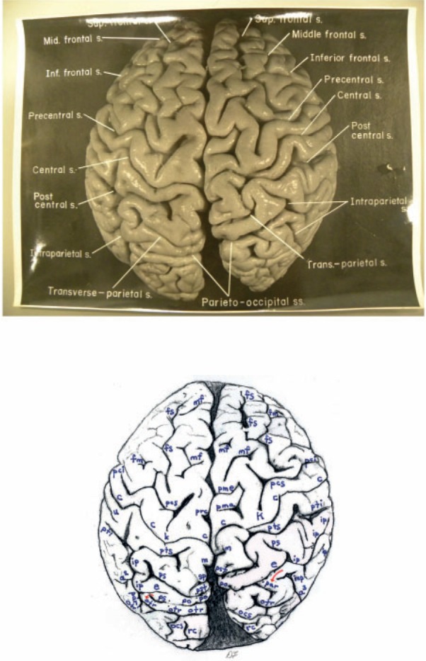
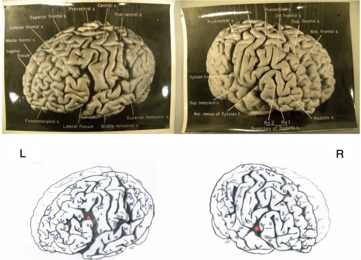
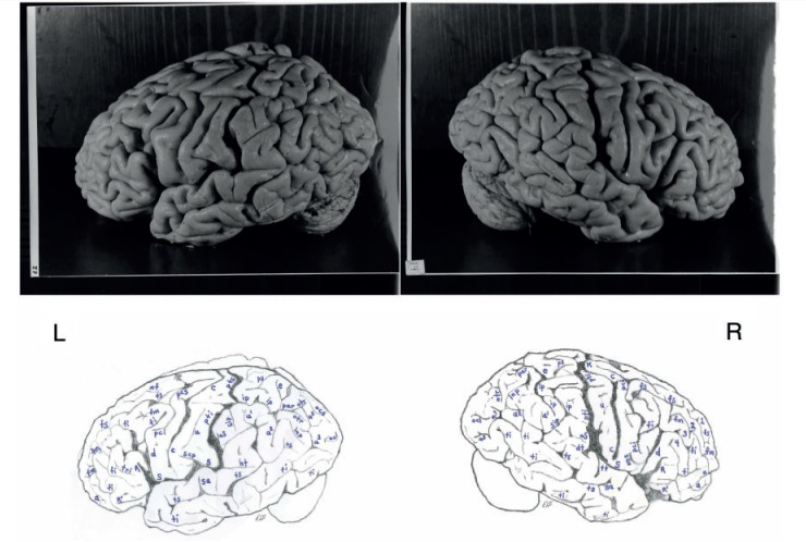
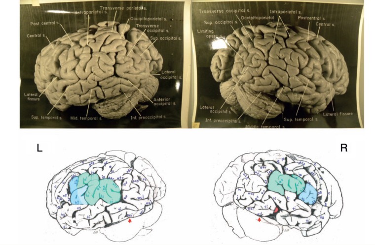
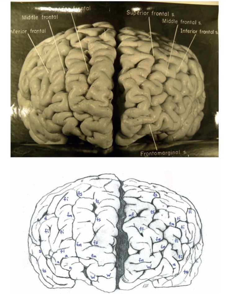
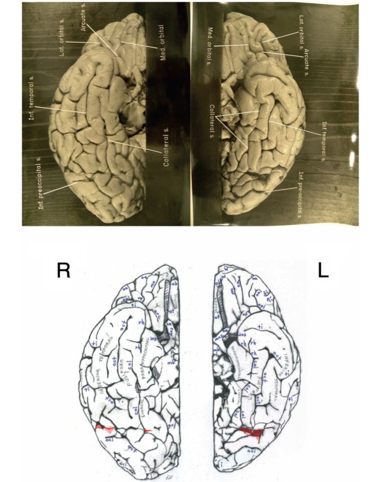
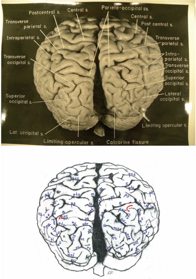
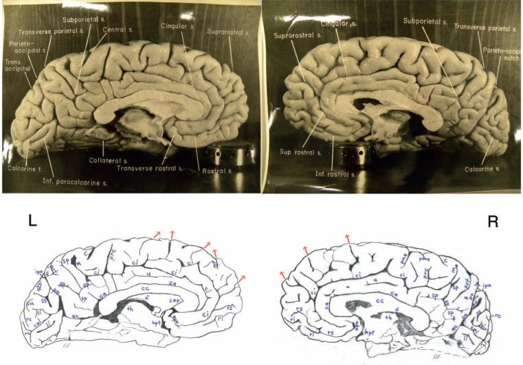
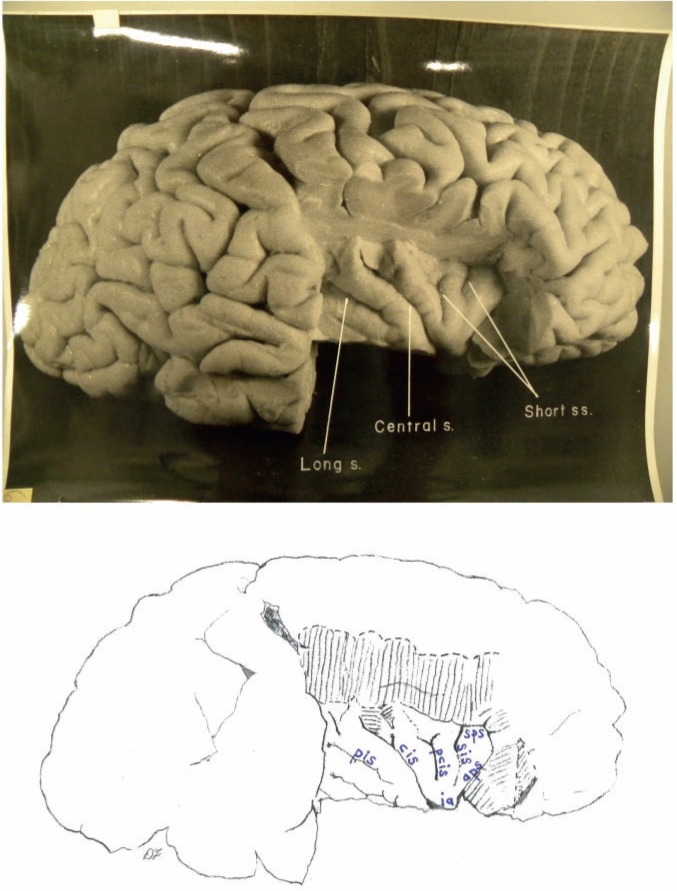
Upon his death in 1955, Albert Einstein's brain was removed, fixed and photographed from multiple angles. It was then sectioned into 240 blocks, and histological slides were prepared. At the time, a roadmap was drawn that illustrates the location within the brain of each block and its associated slides. Here we describe the external gross neuroanatomy of Einstein's entire cerebral cortex from 14 recently discovered photographs, most of which were taken from unconventional angles. Two of the photographs reveal sulcal patterns of the medial surfaces of the hemispheres, and another shows the neuroanatomy of the right (exposed) insula. Most of Einstein's sulci are identified, and sulcal patterns in various parts of the brain are compared with those of 85 human brains that have been described in the literature. To the extent currently possible, unusual features of Einstein's brain are tentatively interpreted in light of what is known about the evolution of higher cognitive processes in humans. As an aid to future investigators, these (and other) features are correlated with blocks on the roadmap (and therefore histological slides). Einstein's brain has an extraordinary prefrontal cortex, which may have contributed to the neurological substrates for some of his remarkable cognitive abilities. The primary somatosensory and motor cortices near the regions that typically represent face and tongue are greatly expanded in the left hemisphere. Einstein's parietal lobes are also unusual and may have provided some of the neurological underpinnings for his visuospatial and mathematical skills, as others have hypothesized. Einstein's brain has typical frontal and occipital shape asymmetries (petalias) and grossly asymmetrical inferior and superior parietal lobules. Contrary to the literature, Einstein's brain is not spherical, does not lack parietal opercula and has non-confluent Sylvian and inferior postcentral sulci.


Figures

Figure 1
Figure 1
Top: Dorsal photograph of Einstein’s brain with original labels. Bottom: Our identifications. a2 = angular; a3 = anterior occipital; c = central; e = processus acuminis; fm = midfrontal; fs = superior frontal; inp = intermediate posterior parietal; ip = intraparietal; m = marginal; mf = medial frontal; ocs = superior occipital; otr = transverse occipital; par = paroccipital; pci = precentral inferior; pcs = precentral superior; pma = marginal precentral; pme = medial precentral; po = parieto-occipital; prc = paracentral; ps = superior parietal; pst = transverse parietal; pti = postcentral inferior; pts = postcentral superior; rc = retrocalcarine; u = unnamed. k = presumed motor cortex for right hand; K = ‘knob’ representing motor cortex for left hand. In both hemispheres, e limits anteriorly the first annectant gyrus, a pli de passage of Gratiolet that connects the parietal and occipital lobes, indicated by red arrows (see also Fig. 7). This figure is reproduced with permission from the National Museum of Health and Medicine.
Figure 2
Figure 2
Top: Photographs of the left (L) and right (R) lateral surfaces of Einstein’s brain taken with the front of the brain rotated toward the viewer, with original labels. Bottom: Our identifications. Numbers 1–4 indicate four gyri in Einstein’s right frontal lobe, rather than three as is typical; K = ‘knob’ representing motor cortex for left hand. Submerged gyri are shaded red near the diagonal sulcus on each side. It is clear from the left hemisphere that the posterior ascending limb of the Sylvian fissure and the postcentral inferior sulcus are not confluent, contrary to the literature. Sulci: a = additional inferior frontal; a1 = ascending branch of the superior temporal sulcus; a2 = angular; aS = posterior ascending limb of the Sylvian; c = central; d = diagonal; dt = descending terminal branch of the Sylvian; fi = inferior frontal; fm = midfrontal; fs = superior frontal; ht = posterior terminal horizontal branch of the Sylvian; ip = intraparietal; mf = medial frontal; pci = precentral inferior; pcs = precentral superior; pma = marginal precentral; pti = postcentral inferior; pts = postcentral superior; R = ascending ramus of anterior Sylvian fissure; R’ = horizontal ramus of anterior Sylvian fissure; S = Sylvian fissure; sa = sulcus acousticus; sca = subcentral anterior; scp = subcentral posterior; sip = intermedius primus of Jensen; ti = inferior temporal; tri = triangular; ts = superior temporal; tt = transverse temporal; u = unnamed; W = fronto-marginal of Wernicke. 1 = superior frontal gyrus; 2 = atypical superior middle frontal gyrus; 3 = atypical inferior middle frontal gyrus; 4 = inferior frontal gyrus (usually the ‘inferior third frontal gyrus’). The figure reproduced with permission from the National Museum of Health and Medicine.
Figure 3
Figure 3
Top: Photographs of the left (L) and right (R) lateral surfaces of Einstein’s brain taken from a traditional view, which lack original labels. Bottom: Our identifications. Numbers 1–4 on the right hemisphere indicate four gyri in Einstein’s right frontal lobe, rather than three as is typical. Sulci: a = additional inferior frontal; a1 = ascending branch of the superior temporal sulcus; a2 = angular; a3 = anterior occipital; aS = posterior ascending limb of the Sylvian; c = central; d = diagonal; dt = descending terminal branch of the Sylvian; e = processus acuminis; fi = inferior frontal; fm = midfrontal; fs = superior frontal; ht = posterior terminal horizontal branch of the Sylvian; inp = intermediate posterior parietal; ip = intraparietal; mf = medial frontal; ocl = lateral occipital; ocs = superior occipital; otr = transverse occipital; par = paroccipital; pci = precentral inferior; pcs = precentral superior; ps = superior parietal; pti = postcentral inferior; pts = postcentral superior; R = ascending ramus of anterior Sylvian fissure; R’ = horizontal ramus of anterior Sylvian fissure; S = Sylvian fissure; sa = sulcus acousticus; sca = subcentral anterior; scp = subcentral posterior; sip = intermedius primus of Jensen; ti = inferior temporal; tri = triangular; ts = superior temporal; tt = transverse temporal; u = unnamed. 1 = superior frontal gyrus; 2 = atypical superior middle frontal gyrus; 3 = atypical inferior middle frontal gyrus; 4 = inferior frontal gyrus (usually the ‘inferior third frontal gyrus’). K = ‘knob’ representing motor cortex for left hand. The figure is reproduced with permission from the National Museum of Health and Medicine.
Figure 4
Figure 4
Top: Photographs of the left (L) and right (R) lateral surfaces of Einstein’s brain taken with the back of the brain rotated towards the viewer, with original labels. Bottom: Our identifications. The arrows indicate the pre-occipital notch at the inferolateral border of each hemisphere, which indicate the approximate inferior boundary between the lateral surfaces of the temporal and occipital lobes; on the right, an apparent artificial cut severed the rostral tip (shaded red) of a gyrus in the posterior part of the inferior temporal lobe. This cut appears to be a lateral extension of that observed on the right side of the base of the brain (Fig. 6). Typically, the supramarginal gyrus surrounds the posterior ascending limb of the Sylvian, and the angular gyrus surrounds the upturned end(s) of superior temporal sulcus. These gyri are separated approximately at the level of the intermedius primus sulcus of Jensen and together form the inferior parietal lobule. The supramarginal gyri are shaded blue; the angular gyri are aqua. In the left hemisphere, part of the cortical region above posterior terminal horizontal branch of the Sylvian is shaded an inbetween colour because it could arguably belong to either gyrus. Einstein’s inferior parietal lobules have different shapes in the two hemispheres, and appear to be relatively larger on the left side. Sulci: a1 = ascending branch of the superior temporal sulcus; a2 = angular; a3 = anterior occipital; aS = posterior ascending limb of the Sylvian; c = central; dt = descending terminal branch of the Sylvian; e = processus acuminis; ht = posterior terminal horizontal branch of the Sylvian; i = inferior polar; inp = intermediate posterior parietal; ip = intraparietal; lc = lateral calcarine; oci = inferior occipital; ocl = lateral occipital; ocs = superior occipital; otr = transverse occipital; par = paroccipital; ps = superior parietal; pti = postcentral inferior; pts = postcentral superior; rc = retrocalcarine; S = Sylvian fissure; scp = subcentral posterior; sip = intermedius primus of Jensen; ti = inferior temporal; ts = superior temporal; u = unnamed. The figure is reproduced with permission from the National Museum of Health and Medicine.
Figure 5
Figure 5
Top: Photograph of a frontal view of Einstein’s brain in an unconventional orientation, with original labels. Bottom: Our identifications of sulci. a = additional inferior frontal; fi = inferior frontal; fm = midfrontal; fs = superior frontal; mf = medial frontal; S = Sylvian fissure; ts = superior temporal; W = fronto-marginal of Wernicke. The figure is reproduced with permission from the National Museum of Health and Medicine.
Figure 6
Figure 6
Top: Separate photographs of the right (R) and left (L) basal views of Einstein’s bisected brain with cerebellum removed and original labels. Bottom: Our identifications. The two photographs are not to the same scale and the right hemisphere is rotated slightly laterally compared with the left, as suggested by a published basal photograph of the entire brain with its cerebellum attached (Witelson et al., 1999b). The base of Einstein’s brain appears to have been accidentally cut, perhaps with a scalpel, as indicated in red shading. This may have occurred during removal of the dura mater (tentorium cerebelli) that separates the dorsum of the cerebellum from the inferior surface of the occipital lobes. Magnifying the photographs on a computer screen should facilitate observation of these cuts. See Fig. 4 for an extension of this cut that reached the right lateral surface of the temporal lobe where it severed the tip of a gyrus (shaded in red). Sulci: arc = arcuate orbital; col = collateral; fi = inferior frontal; i = inferior polar; mo = medial orbital; oa = anterior orbital; oal = lateral anterior orbital; oci = inferior occipital; oct = occipito-temporal; op = posterior orbital; opl = lateral posterior orbital; os = olfactory; R’ = horizontal ramus of anterior Sylvian fissure; rh = rhinal; ti = inferior temporal. Abbreviations of other features: los = lateral olfactory stria; mb = mammillary body; mos = medial olfactory stria; ob = olfactory bulb; on = optic nerve; ot = olfactory tract. The figure is reproduced with permission from the National Museum of Health and Medicine.
Figure 7
Figure 7
Top: Photograph of an occipital view of Einstein’s brain in an unconventional orientation, with original labels. Bottom: Our identifications. In both hemispheres, a processus acuminis limits anteriorly the first annectant gyrus, a pli de passage of Gratiolet that connects the parietal and occipital lobes, indicated by red arrows (see also Fig. 1). See Fig. 10B for shading of the superior and inferior parietal lobules and the occipital lobe on this image. Sulci: a2 = angular; a3 = anterior occipital; c = central; cu = cuneus; e = processus acuminis; inp = intermediate posterior parietal; ip = intraparietal; lc = lateral calcarine; m = marginal; oci = inferior occipital; ocl = lateral occipital; ocs = superior occipital; otr = transverse occipital; par = paroccipital; pcs = precentral superior; po = parieto-occipital; ps = superior parietal; pst = transverse parietal; pti = postcentral inferior; pts = postcentral superior; rc = retrocalcarine; sp = subparietal; ss = superior sagittal; ti = inferior temporal; ts = superior temporal. The figure is reproduced with permission from the National Museum of Health and Medicine.
Figure 8
Figure 8
Top: Photographs of the left (L) and right (R) medial surfaces of Einstein’s brain with original labels. Bottom: Our identifications. Arrows indicate sulci that extend onto the dorsolateral surface of the brain. Sulci: ac = anterior calcarine; apo = anterior parolfactory; c = central; ca = callosal; cal = calcarine; ci = cingulate; cu = cuneus; li = lingual; lp = limiting sulcus of precuneus; m = marginal; mf = medial frontal; otr = transverse occipital; pc = paracalcarine; pma = marginal precentral; pme = medial precentral; po = parieto-occipital; prc = paracentral; pst = transverse parietal; rc = retrocalcarine; ri = inferior rostral; rs = superior rostral; si = inferior sagittal; sp = subparietal; ss = superior sagittal; u = unnamed. Other abbreviations: cc = corpus callosum; f = fornix; hpt = hypothalamus; ipo = parieto-occipital incisure; sep = septum pellucidum; th = thalamus. See text for discussion. The figure is reproduced with permission from the National Museum of Health and Medicine.
Figure 9
Figure 9
Top: Photograph of Einstein’s right insula after removal of the opercula, with original labels. Bottom: Our identifications of sulci: aps = anterior periinsular; cis = central insular; pcis = precentral insular; pis = postcentral insular; sis = short insular; sps = superior periinsular; Other identification: ia = apex of insula. The figure is reproduced with permission from the National Museum of Health and Medicine.
Figure 10
Figure 10
Highlights of Einstein’s brain. (A) Figure 2 of the left lateral surface of Einstein’s brain highlighted to summarize interesting features, which have been darkened. These include a connected precentral superior and inferior sulcus, a long unnamed sulcus in the inferior primary somatosensory cortex, and a posterior ascending limb of the Sylvian fissure that, contrary to the literature, is not confluent with the postcentral inferior sulcus. Unusually expanded primary somatosensory (posterior to the central sulcus) and primary motor cortices (rectangular region below the precentral inferior sulcus) are highlighted in yellow, as are the unusually convoluted surface of the pars triangularis (part of Broca’s speech area) and the frontal polar region. (B) Figure 7 of an occipital view of Einstein’s brain coloured to indicate the approximate boundaries of the superior parietal lobule (purple), inferior parietal lobule (aqua/blue) and occipital lobes (salmon). Presence of four transverse occipital sulci (darkened) is extremely rare, if not unique. Parts of the posterior temporal lobes are uncoloured below the inferior parietal lobules and rostral to the occipital lobes. Although the small striped patch between the superior and inferior parietal lobules on the right belongs with the superior parietal lobule rather than the angular gyrus of the inferior parietal lobule, its relationship with the bordering intraparietal sulcus is usually associated with a location in the angular gyrus. It would therefore be interesting to study the cytoarchitecture of this enigmatic patch of cortex. Notice that the inferior parietal lobule is favoured on the left (and see Fig. 4), while the superior parietal lobule is relatively greater on the right. There is also an asymmetry that favours the right posterior temporal region, and the right occipital lobe is shifted forward relative to the left. (C) Figure 2 of the right lateral surface of Einstein’s brain highlighted to summarize interesting features, including sulci that are darkened. Unusual sulcal patterns include a connected precentral superior and inferior sulcus, a caudal segment of the inferior frontal sulcus that is connected with both the diagonal and precentral inferior sulci, and a long midfrontal sulcus that terminates in the fronto-marginal sulcus of Wernicke. The midfrontal sulcus divides the middle frontal region into two distinct gyri (highlighted in yellow), which causes Einstein’s right frontal lobe to have four rather than the typical three gyri. The enlarged ‘knob’ that probably represents motor cortex for the left hand and the highly convoluted frontal polar region are also highlighted in yellow. (D) Figure 8 of the right medial surface of Einstein’s brain with unusual features highlighted in yellow. The cingulate gyrus has a long unnamed sulcus, the transverse parietal sulcus seems relatively elongated and the cuneus appears to be unusually convoluted. (E) Figure 6 of the basal surface of Einstein’s brain highlighted to show that the left collateral sulcus is divided into two segments, and that part of the fusiform gyrus bridges between these segments to merge with the parahippocampal gyrus. (F) Figure 8 of the left medial surface of Einstein’s brain with unusual features highlighted in yellow. The cingulate gyrus has a long unnamed sulcus, and the cingulate sulcus gives off four inferiorly directed branches (two of which are tiny), which suggest that the cingulate gyrus may be relatively convoluted. The cuneus appears to be unusually convoluted. The figures are reproduced with permission from the National Museum of Health and Medicine.
Figure 11
Figure 11
Part of the original ‘road map’ to the 240 blocks sectioned from Einstein’s brain. A and B correspond with Fig. 2; C and D correspond with Fig. 4, and E with Fig. 5. The figure is reproduced with permission from the National Museum of Health and Medicine.
Figure 12
Figure 12
The remainder of the original ‘road map’ to the 240 blocks sectioned from Einstein’s brain. A–D correspond with Fig. 8. The figure is reproduced with permission from the National Museum of Health and Medicine.

Comment in

References

    1. Abraham C. Possessing genius. New York: St. Martin’s Press; 2002.
    1. Allen JS, Bruss J, Damasio H. Looking for the lunate sulcus: a magnetic resonance imaging study in modern humans. Anat Rec A Discov Mol Cell Evol Biol. 2006;288:867–76. - PubMed
    1. Allman JM, Tetreault NA, Hakeem AY, Manaye KF, Semendeferi K, Erwin JM, et al. The von Economo neurons in frontoinsular and anterior cingulate cortex in great apes and humans. Brain Struct Funct. 2010;214:495–517. - PubMed
    1. Amunts K, Schleicher A, Burgel U, Mohlberg H, Uylings HB, Zilles K. Broca's region revisited: cytoarchitecture and intersubject variability. J Comp Neurol. 1999;412:319–41. - PubMed
    1. Amunts K, Schleicher A, Ditterich A, Zilles K. Broca's region: cytoarchitectonic asymmetry and developmental changes. J Comp Neurol. 2003;465:72–89. - PubMed

Personal name as subject