. 2013 Jan 7;41(1):586-98.
doi: 10.1093/nar/gks1076. Epub 2012 Nov 17.

miRNA repression of translation in vitro takes place during 43S ribosomal scanning

Affiliations

miRNA repression of translation in vitro takes place during 43S ribosomal scanning

Emiliano P Ricci et al. Nucleic Acids Res. .

Abstract

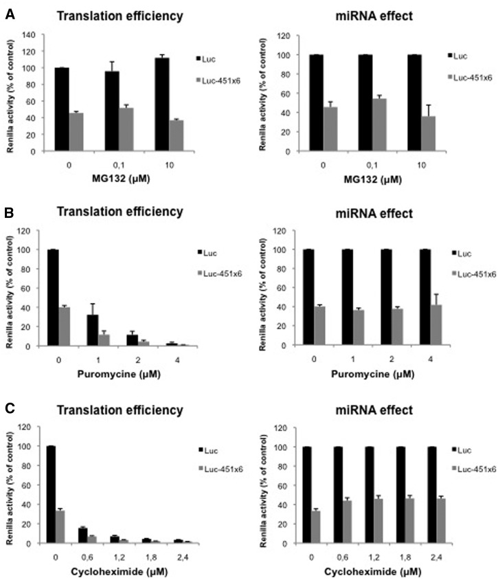
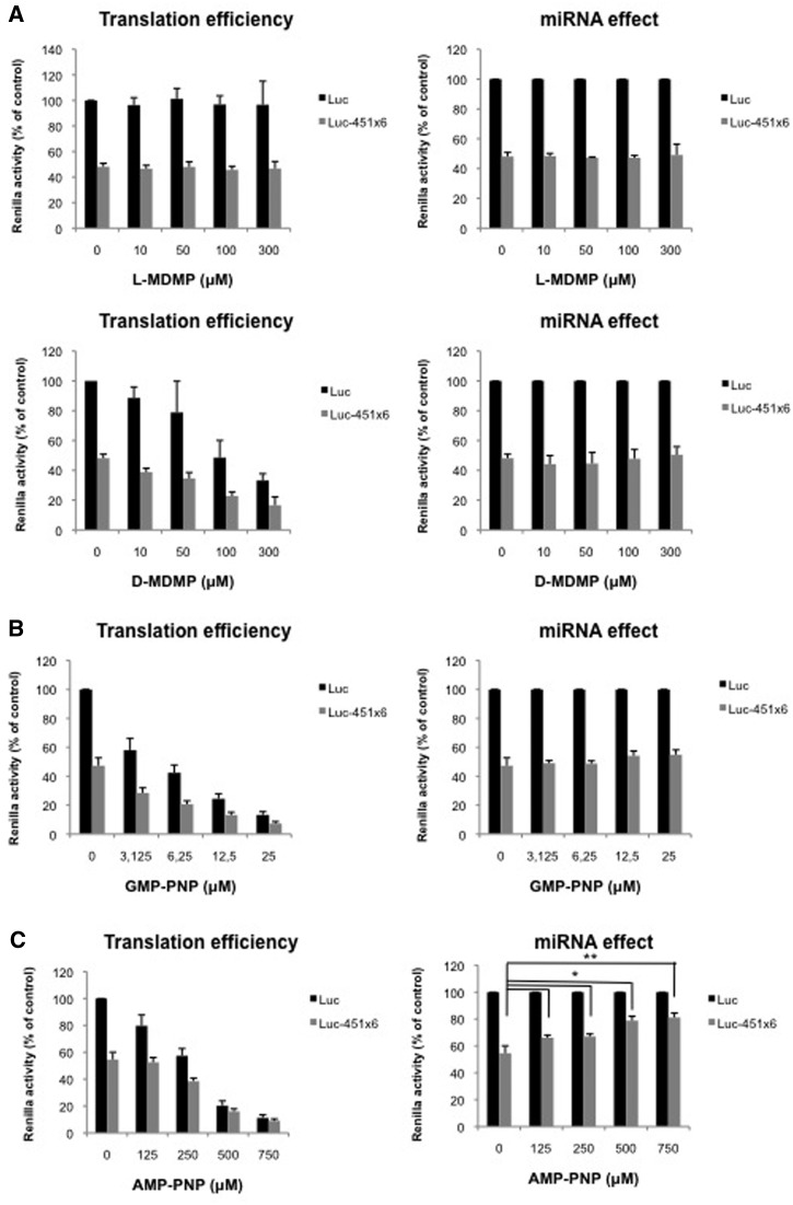
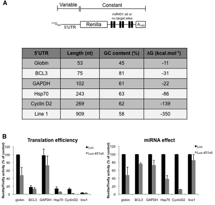
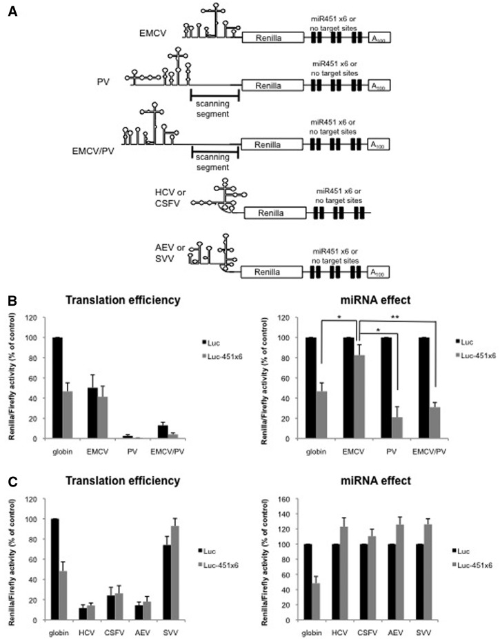
microRNAs (miRNAs) regulate gene expression at multiple levels by repressing translation, stimulating deadenylation and inducing the premature decay of target messenger RNAs (mRNAs). Although the mechanism by which miRNAs repress translation has been widely studied, the precise step targeted and the molecular insights of such repression are still evasive. Here, we have used our newly designed in vitro system, which allows to study miRNA effect on translation independently of deadenylation. By using specific inhibitors of various stages of protein synthesis, we first show that miRNAs target exclusively the early steps of translation with no effect on 60S ribosomal subunit joining, elongation or termination. Then, by using viral proteases and IRES-driven mRNA constructs, we found that translational inhibition takes place during 43S ribosomal scanning and requires both the poly(A) binding protein and eIF4G independently from their physical interaction.


Figures

Figure 1.
Figure 1.
miRNAs do not target translation elongation nor degradation of nascent peptides through the proteasome. (A) Translation of Luc and Luc-451X6 RNAs was carried out in untreated RRL in presence of indicated concentration of MG132. (B) Translation of Luc and Luc-451X6 RNAs was carried out in untreated RRL in presence of indicated concentration of puromycine. (C) Translation of Luc and Luc-451X6 RNAs was carried out in untreated RRL in presence of indicated concentration of cycloheximide. Results are shown as translation efficiency (left panels) and miRNA effect (right panels), as described in ‘Materials and Methods’ section. Error bars correspond to SD obtained from three independent experiments.
Figure 2.
Figure 2.
60S ribosomal joining is not regulated by miRNAs. (A) Translation of Luc and Luc-451X6 RNAs was carried out in untreated RRL in presence of indicated concentration of l-MDMP (top panels) or d-MDMP (bottom panels). (B) Translation of Luc and Luc-451X6 RNAs was carried out in untreated RRL in presence of indicated concentration of GMP-PNP. (C) Translation of Luc and Luc-451X6 RNAs was carried out in untreated RRL in presence of indicated concentration of AMP-PNP. Results are shown as translation efficiency (left panels) and miRNA effect (right panels), as described in ‘Materials and Methods’ section. Error bars correspond to SD obtained from three independent experiments. * corresponds to a P-value <0.05; ** corresponds to a P-value <0.01; (non directional t-test).
Figure 3.
Figure 3.
PABP and eIF4G are required for miRNA-mediated repression independently from their physical interaction. (A) Translation of Luc and Luc-451X6 RNAs was carried out in untreated RRL after addition of HRV 3C protease as indicated. (B) Translation of Luc and Luc-451X6 RNAs was carried out in untreated RRL after addition of 2 μl in vitro translated l-protease, E64 protease inhibitor (10 μM) or both as indicated. (C) Translation of Luc and Luc-451X6 RNAs was carried out in untreated RRL after addition of 2 μl recombinant HIV-2 protease, the palinavir protease inhibitor (10 μM) or both as indicated. (D) Translation of Luc and Luc-451X6 RNAs was carried out in RRL and treated with 2 μl HIV-2 protease for 10 min. The reaction was then stopped by addition of the palinavir protease inhibitor (10 μM) and the recombinant PABP (500 ng) or eIF4G (1 μg) were added to the mixture for a further 50 min before luciferase analysis. Results are shown as translation efficiency (left panels) and miRNA effect (right panels), as described in ‘Materials and Methods’ section. Error bars correspond to SD obtained from three independent experiments; * corresponds to a P-value <0.05; ** corresponds to a P-value <0.01; (non directional t-test).
Figure 4.
Figure 4.
miRNA-mediated translation inhibition is affected by 5′-UTR composition. (A) Schematic representation of the Renilla luciferase RNA used, which contains either no target sites (Luc) or six target sites for miR-451 (Luc-451) at the 3′-end. Expression of this construct was driven by various 5′-UTR that were derived from cellular transcripts as shown on the table (stability of the 5′-UTR was predicted using the mfold program). (B) Translation of Luc and Luc-451X6 RNAs driven by different 5′-UTR was carried out in untreated RRL. Results are shown as translation efficiency (left panel) and miRNA effect (right panel), as described in ‘Materials and Methods’ section. Error bars correspond to SD obtained from three independent experiments.
Figure 5.
Figure 5.
Ribosomal scanning is required for miRNA-mediated inhibition. (A) Schematic representation of the Renilla luciferase RNAs in which translation initiation was driven by EMCV, PV, the EMCV/PV chimera, HCV, CSFV, SVV or AEV IRES. (B) Translation of Luc or Luc-451X6 constructs containing the EMCV, PV, EMCV/PV IRES or globin 5′-UTR as a control, in the untreated RRL. (C) Translation of Luc or Luc-451X6 constructs containing the HCV, CSFV, SVV, AEV IRES or globin 5′-UTR as a control in the untreated RRL. Results are shown as translation efficiency (left panels) and miRNA effect (right panels), as described in ‘Materials and Methods’ section. Error bars correspond to SD obtained from three independent experiments; * corresponds to a P-value <0.05; ** corresponds to a P-value <0.01; (non directional t-test).

References

    1. Bartel DP. MicroRNAs: target recognition and regulatory functions. Cell. 2009;136:215–233. - PMC - PubMed
    1. Friedman RC, Farh KK-H, Burge CB, Bartel DP. Most mammalian mRNAs are conserved targets of microRNAs. Genome Res. 2009;19:92–105. - PMC - PubMed
    1. Bazzini AA, Lee MT, Giraldez AJ. Ribosome profiling shows that miR-430 reduces translation before causing mRNA decay in zebrafish. Science. 2012;336:233–237. - PMC - PubMed
    1. Bethune J, Artus-Revel CG, Filipowicz W. Kinetic analysis reveals successive steps leading to miRNA-mediated silencing in mammalian cells. EMBO Rep. 2012;13:716–723. - PMC - PubMed
    1. Djuranovic S, Nahvi A, Green R. miRNA-mediated gene silencing by translational repression followed by mRNA deadenylation and decay. Science. 2012;336:237–240. - PMC - PubMed

Publication types

MeSH terms