. 2013 Jan 7;41(1):565-74.
doi: 10.1093/nar/gks1074. Epub 2012 Nov 17.

Contribution of intersubunit bridges to the energy barrier of ribosomal translocation

Affiliations

Contribution of intersubunit bridges to the energy barrier of ribosomal translocation

Qi Liu et al. Nucleic Acids Res. .

Abstract

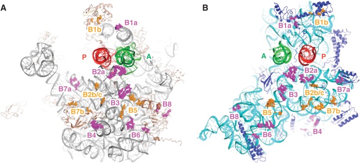
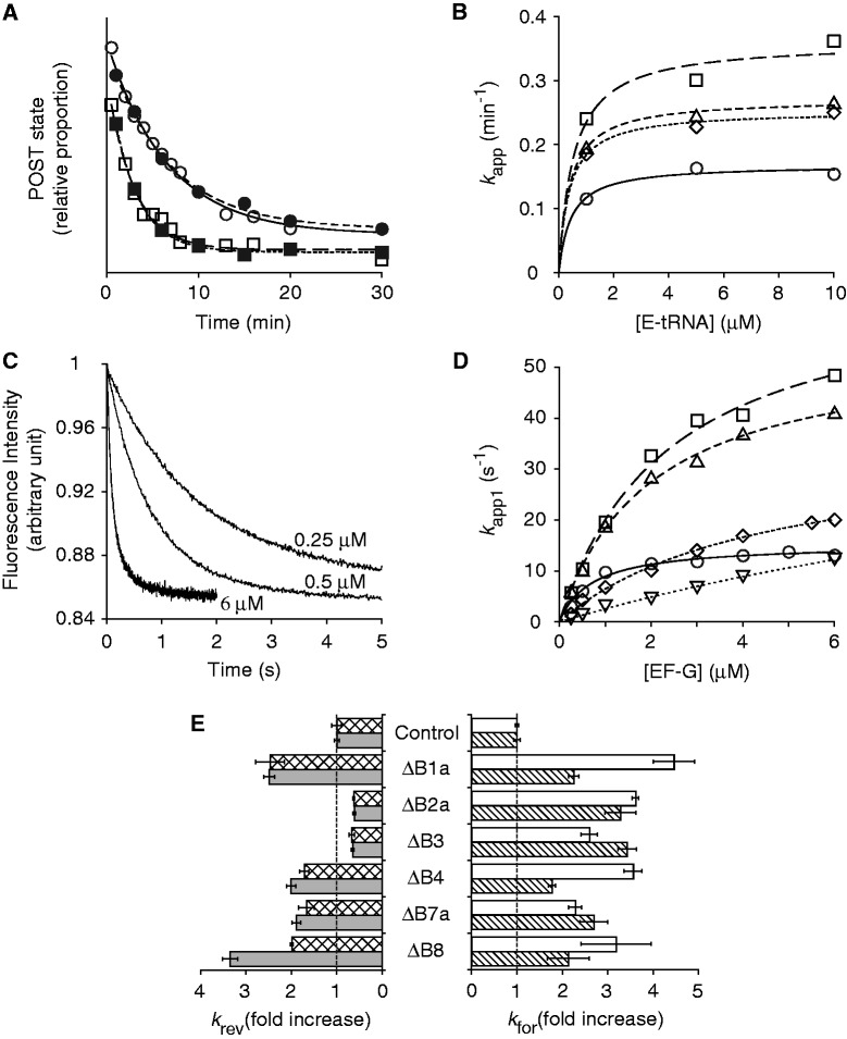
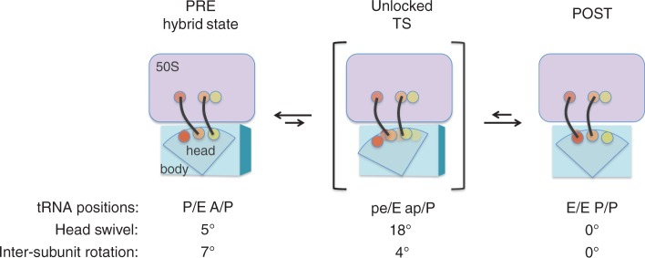
In every round of translation elongation, EF-G catalyzes translocation, the movement of tRNAs (and paired codons) to their adjacent binding sites in the ribosome. Previous kinetic studies have shown that the rate of tRNA-mRNA movement is limited by a conformational change in the ribosome termed 'unlocking'. Although structural studies offer some clues as to what unlocking might entail, the molecular basis of this conformational change remains an open question. In this study, the contribution of intersubunit bridges to the energy barrier of translocation was systematically investigated. Unlike those targeting B2a and B3, mutations that disrupt bridges B1a, B4, B7a and B8 increased the maximal rate of both forward (EF-G dependent) and reverse (spontaneous) translocation. As bridge B1a is predicted to constrain 30S head movement and B4, B7a and B8 are predicted to constrain intersubunit rotation, these data provide evidence that formation of the unlocked (transition) state involves both 30S head movement and intersubunit rotation.


Figures

Figure 1.
Figure 1.
Locations of intersubunit bridges in the ribosome. Components of the bridges formed between the 50S (A) and 30S (B) subunits, viewed from the interface perspective. Bridges targeted in this study, magenta; other bridges, orange; 23S rRNA, gray; 5S rRNA, yellow; 50S proteins, brown; 16S rRNA, cyan; 30S proteins, dark blue; P-site tRNA, red; A-site tRNA, green. This image was generated in PyMOL using PDB entries 2WDG and 2WDI (58).
Figure 2.
Figure 2.
Effects of bridge mutations on translocation. (A) Examples of experiments in which rates of reverse translocation in control (circles) and mutant (ΔB1a; squares) ribosomes were measured, using toeprinting (open symbols) or puromycin-reactivity (closed symbols) assays. Data were fit to a single-exponential function to obtain apparent rates. (B) Apparent rates of reverse translocation (as measured by toeprinting) plotted against E-site tRNA concentration for the control (open circles) and mutant (ΔB1a, open squares; ΔB4, open triangles; ΔB7a, open rhombus) ribosomes. Curves represent hyperbolic fits to the data. (C) Examples of fluorescence traces obtained in measuring rates of forward translocation at different concentrations of EF-G (as indicated). Data were fit to a double-exponential function to obtain apparent rates for the fast (k app1) and slow (k app2) phases. (D) Plots of k app1 versus EF-G concentration for the control (open circles) and a number of mutant ribosomes (ΔB1a, open squares; ΔB4, open triangles; ΔB7a, open rhombus; ΔB8, open inverted triangles). Data were fit to the equation k app = k for•[EF-G]/(K 1/2 + [EF-G]) to yield the parameters shown in Table 2. (E) Summary of the effects of the bridge mutations on the maximal rate of spontaneous reverse translocation [k rev; toeprinting (checkered bars), puromycin-reactivity (gray bars)] and EF-G-catalyzed forward translocation (k for1, white bars; k for2, striped bars).
Figure 3.
Figure 3.
A structural model for translocation. Recent cryo-EM studies by Spahn and coworkers suggest that EF-G can stabilize a conformation of the ribosome in which the 30S head is highly swiveled (18°), while the 30S body/platform is only slightly rotated (4°) with respect to the 50S subunit (53). Such a conformation may resemble the unlocked (transition) state of translocation. 50S, lavender; 30S, blue; A site, yellow; P site, orange; E site, red; tRNAs, black.

Similar articles

Cited by

References

    1. Gavrilova LP, Spirin AS. Stimulation of “non-enzymic” translocation in ribosomes by p-chloromercuribenzoate. FEBS Let. 1971;17:324–326. - PubMed
    1. Pestka S. Studies on the formation of transfer ribonucleic acid-ribosome complexes. VI. Oligopeptide synthesis and translocation on ribosomes in the presence and absence of soluble transfer factors. J. Biol. Chem. 1969;244:1533–1539. - PubMed
    1. Agirrezabala X, Liao HY, Schreiner E, Fu J, Ortiz-Meoz RF, Schulten K, Green R, Frank J. Structural characterization of mRNA-tRNA translocation intermediates. Proc. Natl Acad. Sci. USA. 2012;109:6094–6099. - PMC - PubMed
    1. Chen C, Stevens B, Kaur J, Cabral D, Liu H, Wang Y, Zhang H, Rosenblum G, Smilansky Z, Goldman YE, et al. Single-molecule fluorescence measurements of ribosomal translocation dynamics. Mol. Cell. 2011;42:367–377. - PMC - PubMed
    1. Dunkle JA, Wang L, Feldman MB, Pulk A, Chen VB, Kapral GJ, Noeske J, Richardson JS, Blanchard SC, Cate JH. Structures of the bacterial ribosome in classical and hybrid states of tRNA binding. Science. 2011;332:981–984. - PMC - PubMed

Publication types

MeSH terms