. 2012 Dec;12(12):1627-36.
doi: 10.1002/mabi.201200262. Epub 2012 Nov 19.

In vitro 3D full-thickness skin-equivalent tissue model using silk and collagen biomaterials

Affiliations

In vitro 3D full-thickness skin-equivalent tissue model using silk and collagen biomaterials

Evangelia Bellas et al. Macromol Biosci. 2012 Dec.

Abstract

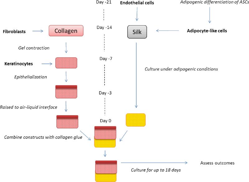
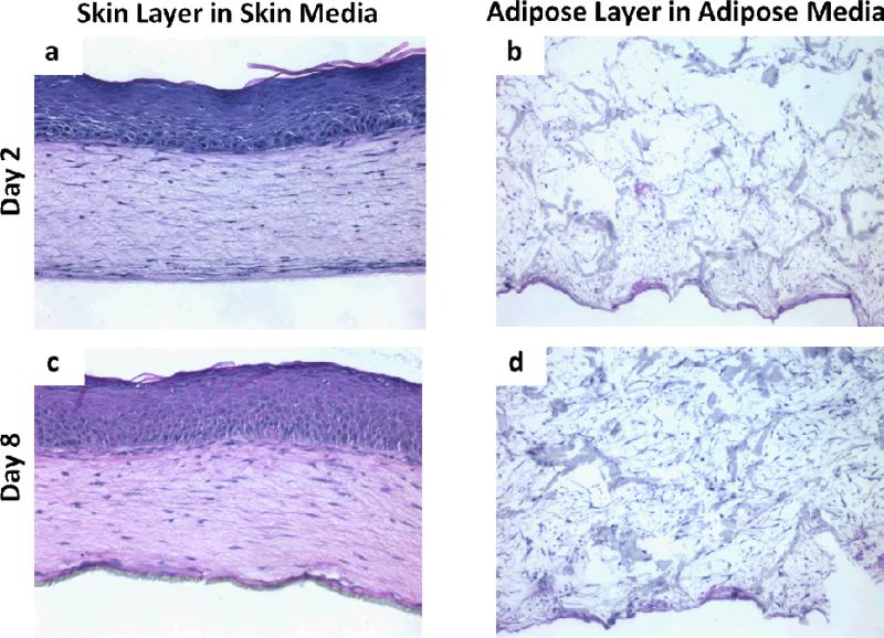
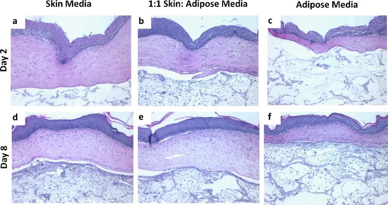
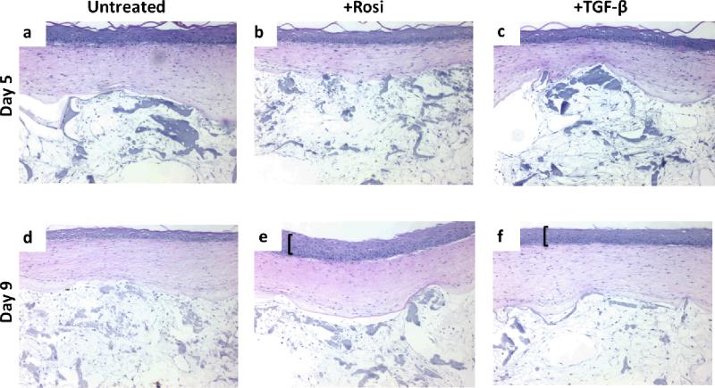
Current approaches to skin equivalents often only include the epidermis and dermis. Here, a full-thickness skin equivalent is described including epidermis, dermis, and hypodermis, that could serve as an in vitro model for studying skin biology or as a platform for consumer product testing. The construct is easy to handle and is maintained for >14 d while expressing physiological morphologies of the epidermis and dermis, seen by keratin 10, collagens I and IV expression. The skin equivalent produces glycerol and leptin, markers of adipose metabolism. This work serves as a foundation for understanding a few necessary factors needed to develop a stable, functional model of full-thickness skin.


Figures

Figure 1
Figure 1
Experimental outline for tri-layer culture. Skin and adipose tissues were cultured independently prior to being combined at ‘day 0’. At day -21, endothelial cells are seeded onto silk sponges, while adipose derived stem cells undergo adipogenic differentiation in 2D. At day 14, the adipocyte-like cells are added to the endothelial seeded silk sponges and the co-cultures were maintained in a 1:1 adipogenic:endothelial cell media. Also at day 14, fibroblasts are cast within a collagen gel. After the cell contracts, keratinocytes are seeded on top of the collagen gel. After epithelialization has occurred the construct was raised to the air-liquid interface for 3 days. On day 0, the 2 constructs were combined with a thin layer of collagen gel as a binder. At this point the tri-layer construct was cultured in a 1:1 mix of the co-culture media to skin media.
Figure 2
Figure 2
H&E images of positive controls for skin and adipose constructs after 2 and 8 days in culture. Skin constructs demonstrated a well-organized, multilayer epithelium by day 2 (a) that demonstrated all 4 morphological layers and a collagen lattice in which resident fibroblasts showed their characteristic spindled morphology. This epithelium was further developed by day 8 (c), where it showed a columnar basal cell layer and more complete development of the spinous cell layer. By day 8, cells populated the entire silk sponge in the adipose construct. Images are representative of n=2 per group.
Figure 3
Figure 3
Constructs cultured in 1:1 skin: adipose media had more physiologically relevant features. H&E images of tri-layer constructs in different media after 2 and 8 days in culture. Images are representative of n=2 per group.
Figure 4
Figure 4
Tri-layer constructs were responsive to various drugs. Rosiglitizone (Rosi), an activator of the adipogenic program, caused hyper-proliferation (bracket) at the basal layer of the epidermis at 9 days. TGF-β, also caused hyper-proliferation, to a lesser extent than the Rosi group, at the basal layer of the epidermis at 9 days. The adipose regions did not appear affected, morphologically, by the drug treatments. Images are representative of n=2 per group.

References

    1. Welss T, Basketter D. a, Schröder KR. In vitro skin irritation: facts and future. State of the art review of mechanisms and models. Toxicology in vitro: an international journal published in association with BIBRA. 2004;18:231–43. - PubMed
    1. Egles C, Garlick JA, Shamis Y. Chapter 24 Three-Dimensional Human Tissue Models of Wounded Skin. Methods in Molecular Biology. :345–359. doi:10.1007/978-1-60761-380-0. - PMC - PubMed
    1. Carlson MW, Alt-holland A, Egles C, Garlick JA. Three-Dimensional Tissue Models of Normal and Diseased Skin. 2008:1–17. doi:10.1002/0471143030.cb1909s41. - PMC - PubMed
    1. Metcalfe AD, Ferguson MWJ. Tissue engineering of replacement skin: the crossroads of biomaterials, wound healing, embryonic development, stem cells and regeneration. Journal of the Royal Society, Interface / the Royal Society. 2007:413–37. - PMC - PubMed
    1. Trottier V, Marceau-Fortier G, Germain L, Vincent C, Fradette J. IFATS collection: Using human adipose-derived stem/stromal cells for the production of new skin substitutes. Stem cells. 2008;26:2713–23. - PubMed

Publication types