

Adenocarcinoma cells in effusion cytology as a diagnostic pitfall with potential impact on clinical management: a case report with brief review of immunomarkers
- PMID: 23161830
- PMCID: PMC7682751
- DOI: 10.1002/dc.22915
Adenocarcinoma cells in effusion cytology as a diagnostic pitfall with potential impact on clinical management: a case report with brief review of immunomarkers
Abstract
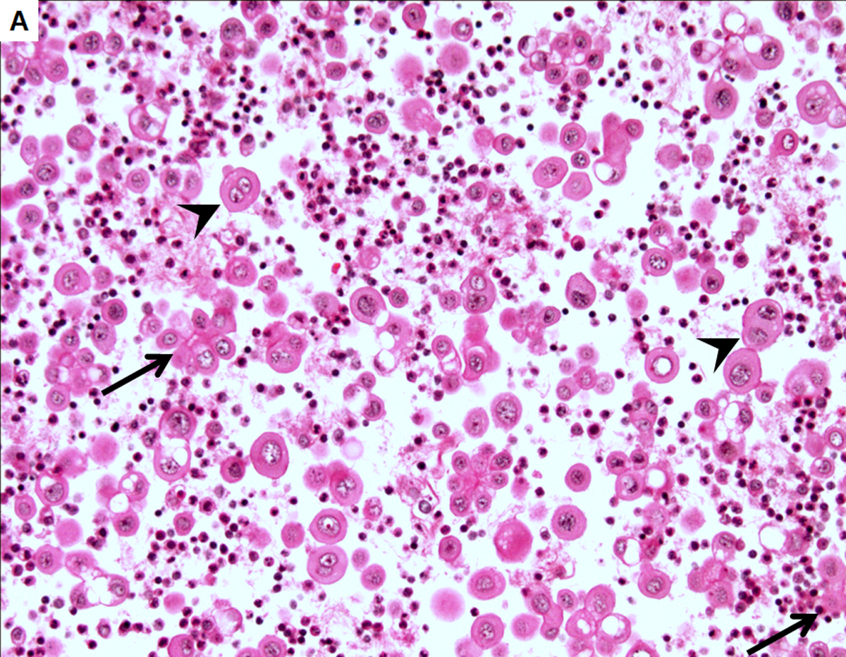
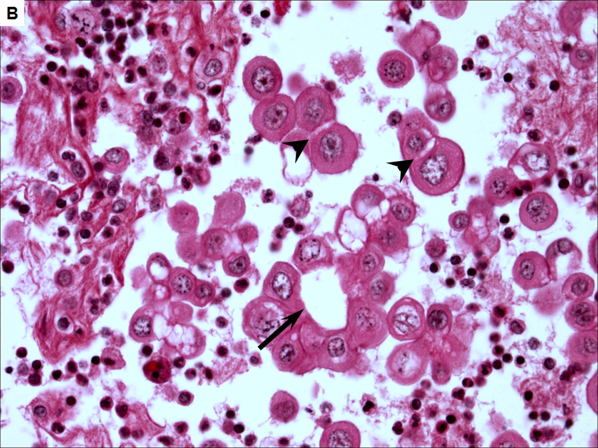
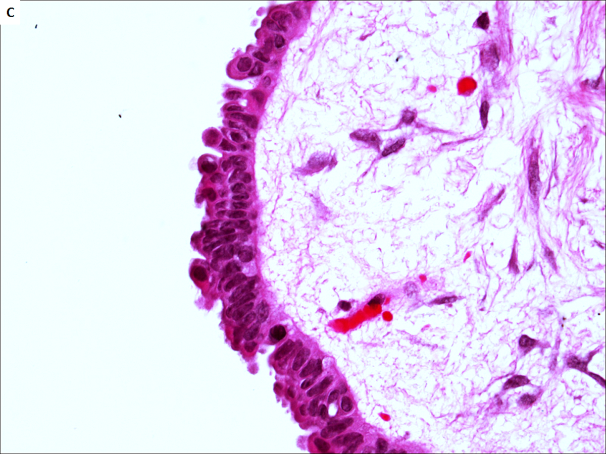
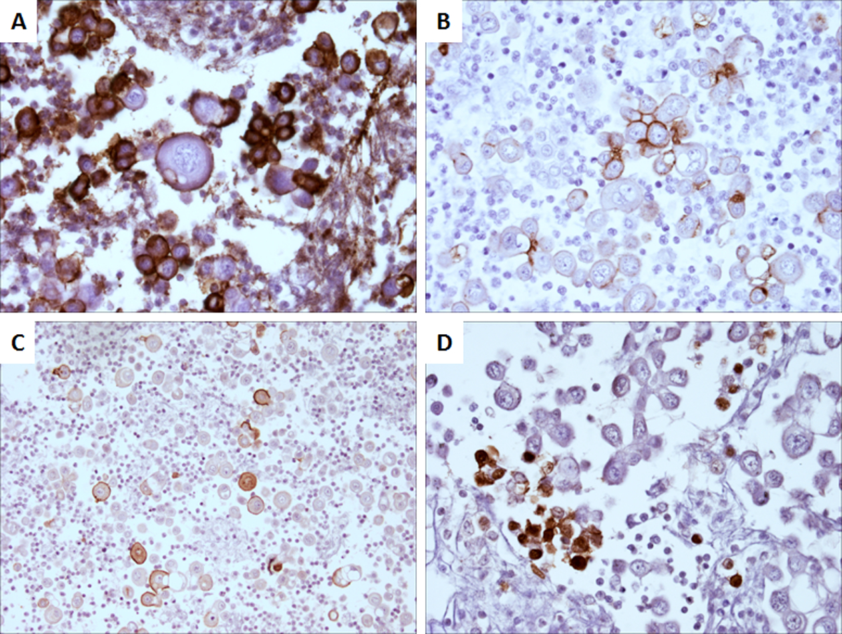
Distinguishing metastatic carcinoma cells from reactive mesothelial cells in effusion samples is often challenging based on morphology alone. Metastatic carcinoma cells in fluid samples may mimic reactive mesothelial cells due to overlapping cytological features. We report a case of a pleural effusion in a 51-year-old female patient with a medical history significant for bilateral ovarian tumors and peritoneal implants diagnosed as serous tumor of borderline malignant potential. The effusion was composed almost entirely of adenocarcinoma cells that morphologically mimicked reactive mesothelial cells. The diagnosis of metastatic adenocarcinoma was made after a wide immunostaining panel of antibodies. Recognizing metastatic adenocarcinoma cells in effusion samples can be challenging and an accurate diagnosis may have significant impact on clinical management as demonstrated by this case.
Keywords: adenocarcinoma cells; cytology; effusion; immunostaining; reactive mesothelial cells.
Copyright © 2012 Wiley Periodicals, Inc., a Wiley company.
Figures
References
-
- Hausheer FH, Yarbro JW. Diagnosis and treatment of malignant pleural effusion. Cancer Metastasis Rev 1987; 6:23–40. - PubMed
-
- DeMay RM. The art & science of cytopathology, Vol., ASCP Press: Chicago, 1996.
-
- Fetsch PA, Abati A. Immunocytochemistry in effusion cytology: a contemporary review. Cancer 2001; 93:293–308. - PubMed
-
- Ordonez NG. Role of immunohistochemistry in differentiating epithelial mesothelioma from adenocarcinoma. Review and update. Am J Clin Pathol 1999; 112:75–89. - PubMed
-
- Ordonez NG. Value of immunohistochemistry in distinguishing peritoneal mesothelioma from serous carcinoma of the ovary and peritoneum: a review and update. Adv Anat Pathol 2006; 13:16–25. - PubMed
Publication types
MeSH terms
Substances
Grants and funding
LinkOut - more resources
Full Text Sources
Medical
