Raman spectroscopy in biomedicine - non-invasive in vitro analysis of cells and extracellular matrix components in tissues
- PMID: 23161832
- PMCID: PMC3644878
- DOI: 10.1002/biot.201200163
Raman spectroscopy in biomedicine - non-invasive in vitro analysis of cells and extracellular matrix components in tissues
Abstract
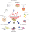
Raman spectroscopy is an established laser-based technology for the quality assurance of pharmaceutical products. Over the past few years, Raman spectroscopy has become a powerful diagnostic tool in the life sciences. Raman spectra allow assessment of the overall molecular constitution of biological samples, based on specific signals from proteins, nucleic acids, lipids, carbohydrates, and inorganic crystals. Measurements are non-invasive and do not require sample processing, making Raman spectroscopy a reliable and robust method with numerous applications in biomedicine. Moreover, Raman spectroscopy allows the highly sensitive discrimination of bacteria. Rama spectra retain information on continuous metabolic processes and kinetics such as lipid storage and recombinant protein production. Raman spectra are specific for each cell type and provide additional information on cell viability, differentiation status, and tumorigenicity. In tissues, Raman spectroscopy can detect major extracellular matrix components and their secondary structures. Furthermore, the non-invasive characterization of healthy and pathological tissues as well as quality control and process monitoring of in vitro-engineered matrix is possible. This review provides comprehensive insight to the current progress in expanding the applicability of Raman spectroscopy for the characterization of living cells and tissues, and serves as a good reference point for those starting in the field.
Copyright © 2013 WILEY-VCH Verlag GmbH & Co. KGaA, Weinheim.
Figures
Similar articles
-
Multivariate analysis of Raman spectra for discriminating human collagens: In vitro identification of extracellular matrix collagens produced by an osteosarcoma cell line.Spectrochim Acta A Mol Biomol Spectrosc. 2025 Mar 5;328:125434. doi: 10.1016/j.saa.2024.125434. Epub 2024 Nov 14. Spectrochim Acta A Mol Biomol Spectrosc. 2025. PMID: 39612534
-
Non-contact, label-free monitoring of cells and extracellular matrix using Raman spectroscopy.J Vis Exp. 2012 May 29;(63):3977. doi: 10.3791/3977. J Vis Exp. 2012. PMID: 22688496 Free PMC article.
-
Non-invasive analysis of cell cycle dynamics in single living cells with Raman micro-spectroscopy.J Cell Biochem. 2008 Jul 1;104(4):1427-38. doi: 10.1002/jcb.21720. J Cell Biochem. 2008. PMID: 18348254
-
Single cell Raman spectroscopy for cell sorting and imaging.Curr Opin Biotechnol. 2012 Feb;23(1):56-63. doi: 10.1016/j.copbio.2011.11.019. Epub 2011 Dec 2. Curr Opin Biotechnol. 2012. PMID: 22138495 Review.
-
Raman spectroscopy: A promising analytical tool used in human reproductive medicine.J Pharm Biomed Anal. 2024 Oct 15;249:116366. doi: 10.1016/j.jpba.2024.116366. Epub 2024 Jul 15. J Pharm Biomed Anal. 2024. PMID: 39029353 Review.
Cited by
-
Generation and Assessment of Functional Biomaterial Scaffolds for Applications in Cardiovascular Tissue Engineering and Regenerative Medicine.Adv Healthc Mater. 2015 Nov 18;4(16):2326-41. doi: 10.1002/adhm.201400762. Epub 2015 Mar 16. Adv Healthc Mater. 2015. PMID: 25778713 Free PMC article. Review.
-
Compositional assessment of bone by Raman spectroscopy.Analyst. 2021 Dec 6;146(24):7464-7490. doi: 10.1039/d1an01560e. Analyst. 2021. PMID: 34786574 Free PMC article. Review.
-
Raman spectrum: A potential biomarker for embryo assessment during in vitro fertilization.Exp Ther Med. 2017 May;13(5):1789-1792. doi: 10.3892/etm.2017.4160. Epub 2017 Feb 23. Exp Ther Med. 2017. PMID: 28565768 Free PMC article.
-
Candida parapsilosis biofilm identification by Raman spectroscopy.Int J Mol Sci. 2014 Dec 22;15(12):23924-35. doi: 10.3390/ijms151223924. Int J Mol Sci. 2014. PMID: 25535081 Free PMC article.
-
Non-invasive detection of DNA methylation states in carcinoma and pluripotent stem cells using Raman microspectroscopy and imaging.Sci Rep. 2019 May 7;9(1):7014. doi: 10.1038/s41598-019-43520-z. Sci Rep. 2019. PMID: 31065074 Free PMC article.
References
-
- Votteler M, Carvajal Berrio DA, Pudlas M, Walles H, et al. Non-contact, label-free monitoring of cells and extracellular matrix using raman spectroscopy. J. Vis. Exp. 2012;63:e3977. doi: 10.3791/3977. - DOI - PMC - PubMed
-
- Raman CV, Krishnan KS. A new type of secondary radiation. Nature. 1928;501–505:121.
-
- LaPlant, F., Lasers, spectrographs, and detectors in: Matousek, P., Morris, M. D. (Eds.), Emerging Raman Applications and Techniques in Biomedical and Pharmaceutical FieldsSpringer Berlin, Heidelberg 2010, pp. 1–24.
-
- Mariani MM, Deckert V. Raman spectroscopy: Principles, benefits & applications. Bunsen-Magazin. 2012;14:136–147.
Publication types
MeSH terms
LinkOut - more resources
Full Text Sources
Miscellaneous


