Randomized Controlled Trial
. 2013 May;27(4):381-4.
doi: 10.1177/1545968312465193. Epub 2012 Nov 16.

Motor representations in the intact hemisphere of the rat are reduced after repetitive training of the impaired forelimb

Affiliations
Randomized Controlled Trial

Motor representations in the intact hemisphere of the rat are reduced after repetitive training of the impaired forelimb

Scott Barbay et al. Neurorehabil Neural Repair. 2013 May.

Abstract

Background: During recovery from a unilateral cortical stroke, spared cortical motor areas in the contralateral (intact) cerebral cortex are recruited. Preclinical studies have demonstrated that compensation with the less-impaired limb may have a detrimental inhibitory effect on the intact cortical hemisphere and could impede recovery of the more-impaired limb. However, evidence from detailed neurophysiological mapping studies in animal models is lacking.

Objectives: The present study examines neurophysiological changes in the intact hemisphere of the rat following a unilateral ischemic infarct to cortical forelimb motor areas.

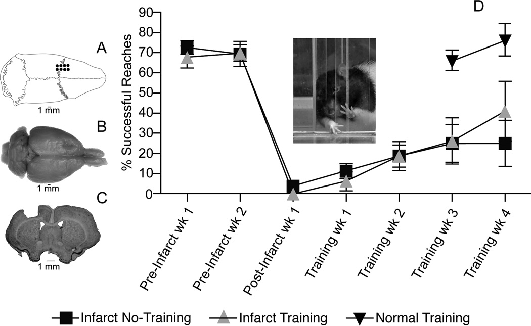
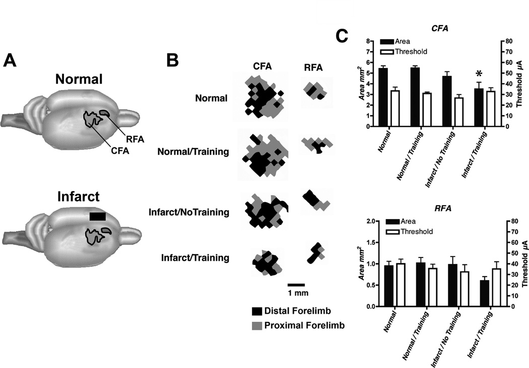
Methods: A total of 8 rats were trained for 2 weeks on a reach and retrieval task prior to an ischemic infarct induced by the vasoconstrictor endothelin-1 injected into the cortical gray matter encompassing the 2 forelimb motor representations: the caudal forelimb area (CFA) and the rostral forelimb area (RFA). Animals were randomly assigned to an infarct/training group (n = 4) or an infarct/no-training group (ie, spontaneous recovery, n = 4). After a 5-week postinfarct period, motor areas of the intact hemisphere (CFA and RFA) were characterized using intracortical microstimulation techniques. The resulting maps of evoked movements were compared with maps derived from CFA and RFA in normal rats (normal, n = 5; normal/training, n = 4).

Results: Compared with the normal/no-training group, CFA representations were significantly smaller in the infarct/training group but not in the infarct/no-training group. No significant differences were found in RFA.

Conclusions: Repetitive training of the more-impaired forelimb during the postinfarct recovery period reduces the size of motor representations in the intact hemisphere.


Figures

Figure 1
Figure 1
Motor performance after ET-1 lesion in motor cortex. A. Dorsal view of skull showing location of boreholes for ET-1 injections. B. Dorsal view of fixed brain showing location of ET-1 lesion in left motor cortex. C. Coronal section through the level of the caudal forelimb area (CFA) at bregma showing ET-1 lesion. Lesion extended through all cortical layers, but did not invade the underlying white matter. Lesion volume was derived from coronal sections imported into NIH Image J software and estimated by the Cavalieri method: Infarct/Training Group = 17.35±0.25 mm3; Infarct/No Training Group = 17.04±0.49 mm3 (p>0.05). Lesion volumes are similar to those predicted based on distribution of ET-1 injections (16mm3). D. Motor performance on single-pellet retrieval task. Rats were trained daily to reach through an opening in a Plexiglas chamber to retrieve small 45 mg food pellets (Bioserve, Frenchtown, NJ) located on a shelf placed above the floor outside the chamber (see inset). Once forelimb preference for unrestricted reaching was established, a movable wall inside the chamber allowed reaching with only the right or left forelimb. Food pellets were delivered one at a time for 60 trials during each session of training. A trial ended with a successful reach and retrieval to the rat’s mouth, after an unsuccessful retrieval when a pellet was contacted but not grasped, or after five failed reach attempts without contacting the pellet. A training session ended after 20 minutes regardless of the number of trials completed. Postinfarct training was conducted daily for 28 days; training for the normal group was conducted daily for 14 days (see text). Performance was assessed once per week during two weeks of baseline training, 1 week after the infarct prior to rehabilitative training and once per week after rehabilitative training.
Figure 2
Figure 2
Neurophysiological maps of the caudal and rostral forelimb areas (CFA and RFA, respectively) in the contralateral, intact hemisphere after an ET-1 lesion in motor cortex. An ICMS stimulus was delivered as a train burst of 13, 0.2 ms cathodal, monophasic pulses, delivered at 350 Hz by a constant-current stimulator (Model BSI-2, BAK Electronics Inc.) at a rate of one train per second. High-resolution motor maps were derived in both CFA and RFA (350µm resolution). A. Location of CFA and RFA motor maps. B. Representational maps of CFA and RFA were delineated with customized software and analyzed quantitatively with NIH Image software. Representative maps from each group are displayed. Shown are maps derived from the right hemisphere of rats with a right forelimb preference. Distal forelimb is shown in black and proximal forelimb is shown in grey. C. Double-Y plot showing map areas and movement thresholds. Areal measurements (Mean ± SEM) of CFA and RFA in intact and infarcted rats are presented on the left y-axis. Current thresholds (Mean ± SEM) for evoking a forelimb movement in CFA and RFA are presented on the right y-axis. There were no significant threshold differences among the four groups in CFA or RFA. Only the Infarct/Training group had a significantly smaller CFA map relative to normal rats. Asterisk = p<0.05.

Similar articles

Cited by

References

    1. Bestmann S, Swayne O, Blankenburg F, et al. The role of contralesional dorsal premotor cortex after stroke as studied with concurrent TMS-fMRI. J Neurosci. 2010 Sep 8;30(36):11926–11937. - PMC - PubMed
    1. Adkins DL, Voorhies AC, Jones TA. Behavioral and neuroplastic effects of focal endothelin-1 induced sensorimotor cortex lesions. Neuroscience. 2004;128(3):473–486. - PubMed
    1. Napieralski JA, Butler AK, Chesselet MF. Anatomical and functional evidence for lesion-specific sprouting of corticostriatal input in the adult rat. J Comp Neurol. 1996 Sep 30;373(4):484–497. - PubMed
    1. Nishibe M, Barbay S, Guggenmos D, Nudo RJ. Reorganization of motor cortex after controlled cortical impact in rats and implications for functional recovery. J. Neurotrauma. 2010 Dec;27(12):2221–2232. - PMC - PubMed
    1. Allred RP, Cappellini CH, Jones TA. The "good" limb makes the "bad" limb worse: experience-dependent interhemispheric disruption of functional outcome after cortical infarcts in rats. Behav Neurosci. 2010 Feb;124(1):124–132. - PMC - PubMed

Publication types

MeSH terms