Comparative Study
. 2013 Jan 15;304(2):H246-52.
doi: 10.1152/ajpheart.00129.2012. Epub 2012 Nov 16.

Cardiomyocyte architectural plasticity in fetal, neonatal, and adult pig hearts delineated with diffusion tensor MRI

Affiliations
Comparative Study

Cardiomyocyte architectural plasticity in fetal, neonatal, and adult pig hearts delineated with diffusion tensor MRI

Lei Zhang et al. Am J Physiol Heart Circ Physiol. .

Abstract

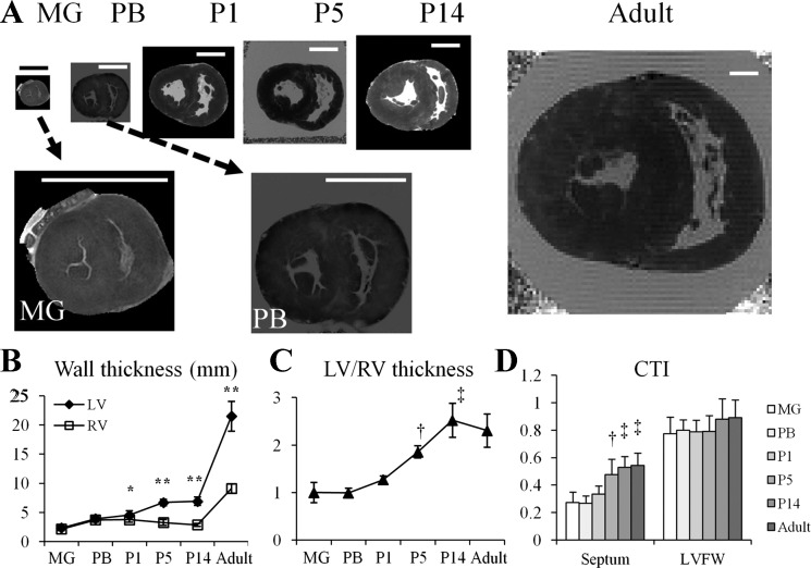
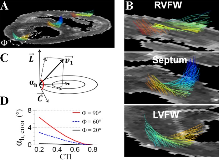
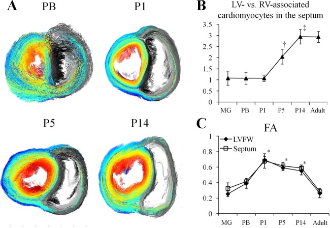
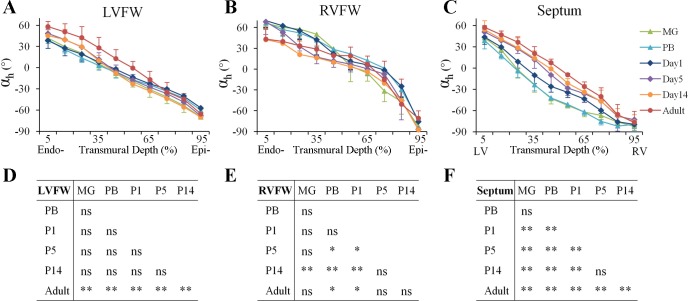
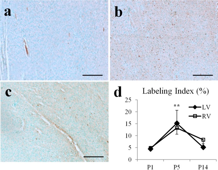
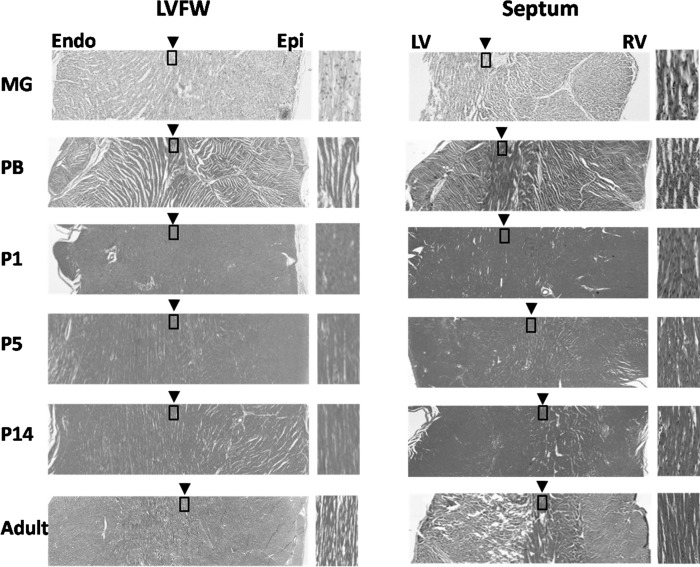
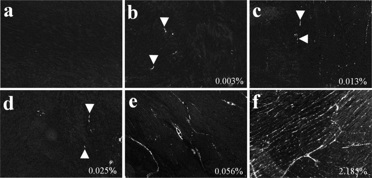
Cardiomyocyte organization is a critical determinant of coordinated cardiac contractile function. Because of the acute opening of the pulmonary circulation, the relative workload of the left ventricle (LV) and right ventricle (RV) changes substantially immediately after birth. We hypothesized that three-dimensional cardiomyocyte architecture might be required to adapt rapidly to accommodate programmed perinatal changes of cardiac function. Isolated fixed hearts from pig fetuses or pigs at midgestation, preborn, postnatal day 1 (P1), postnatal day 5, postnatal day 14 (P14), and adulthood (n = 5 for each group) were acquired for diffusion-weighted magnetic resonance imaging. Cardiomyocyte architecture was visualized by three-dimensional fiber tracking and was quantitatively evaluated by the measured helix angle (α(h)). Upon the completion of MRI, hearts were sectioned and stained with hematoxylin/eosin (H&E) to evaluate cardiomyocyte alignment, with picrosirius red to evaluate collagen content, and with anti-Ki67 to evaluate postnatal cell proliferation. The helical architecture of cardiomyocyte was observed as early as the midgestational period. Postnatal changes of cardiomyocyte architecture were observed from P1 to P14, which primary occurred in the septum and RV free wall (RVFW). In the septum, the volume ratio of LV- vs. RV-associated cardiomyocytes rapidly changed from RV-LV balanced pattern at birth to LV dominant pattern by P14. In the RVFW, subendocardial α(h) decreased by ~30° from P1 to P14. These findings indicate that the helical architecture of cardiomyocyte is developed as early as the midgestation period. Substantial and rapid adaptive changes in cardiac microarchitecture suggested considerable developmental plasticity of cardiomyocyte form and function in the postnatal period in response to altered cardiac mechanical function.


Figures

Fig. 1.
Fig. 1.
Developmental changes of the pig heart morphology. A: representative short-axis images of pig heart at midgestation (MG); preborn (PB); postnatal day 1, postnatal day 5, and postnatal day 14 (P1, P5, and P14, respectively); and adult. Scale bar = 1 cm. B: wall thickness of left ventricle (LV) free wall (LVFW) and right ventricle (RV) free wall (RVFW) in pig hearts at different ages. Thickness of the LVFW increased more rapidly than that of RVFW in newborn hearts. *P < 0.05 and **P < 0.001 compared with the thickness of RVFW at the same timepoint. C: thickness ratio of LVFW to RVFW increased steadily from P1 to P14. †P < 0.05 compared with MG, PB, or P1; ‡P < 0.05 compared with MG, PB, P1, or P5. D: curvature thickness index (CTI) of the septum and LVFW. After birth, CTI of the septum rapidly increased from P1 to P5. †P < 0.05 compared with MG or PB; ‡P < 0.05 compared with MG, PB, or P1.
Fig. 2.
Fig. 2.
The 3-dimensional organization of cardiomoycytes. A and B: representative image of 3-dimensional cardiomyocyte orientation on a short-axis slice of P1 pig heart shows the transmural shift of cardiomyocyte orientation in the RVFW, septum, and LVFW. A Φ = 20° sector at the center of LVFW was selected for quantitatively analysis of αh. C: definition of αh as the angle between circumferential orientation (C) and the projection of cardiomyocyte orientation (v1) on the tangential plane, which is defined by the circumferential and longitudinal (L) axis. D: maximal measurement error in αhh, error) under different CTIs and within different sizes of regions of interest. When a region of interest was selected at a Φ = 20° sector, measurement error in αh was <1° when CTI changed from 0.2 to 0.8.
Fig. 3.
Fig. 3.
Volume ratio of LV- vs. RV-associated cardiomyocytes in the septum. A: representative projection views of cardiomyocyte architecture showed decreased fraction of RV-associated cardiomyocytes (gray color) in the septum from P1 to P14. The LV-associated cardiomyocytes were color-coded with its αh on a midventricular short-axis slice, which shows those cardiomoycytes extended from LV to RV. B: volume ratio of LV- vs. RV-associated cardiomyocytes in the septum increased ∼2-fold from P1 to P14. †P < 0.05 compared with MG, PB, or P1; ‡P < 0.05 compared with MG, PB, P1, or P5. C: fractional anisotropy (FA) of water diffusivity in P1, P5, and P14 hearts were comparable (P = not significant for all comparisons), but all higher than MG, PB, and adult hearts (*P < 0.05 compared with MG, PB, or adult).
Fig. 4.
Fig. 4.
Transmural distribution of αh in the LVFW, RVFW, and septum. AC: transmural distribution of αh in the LVFW, RVFW, and septum. In LVFW and RVFW, data were analyzed from the endocardial (Endo-) surface (5% wall depth) to the epicardial (Epi-) surface (95% wall depth). In the septum, data were analyzed from the LV endocardial surface (5%) to the RV endocardial surface (95%). D-F: post hoc multiple comparisons of transmural distribution of αh between different age groups in the LVFW, RVFW, and septum. *P < 0.05; **P < 0.001. ns, not significant.
Fig. 5.
Fig. 5.
Anti-Ki67 staining of cell proliferation in pig hearts. AC: representative images of stained Ki67-positive cells (in brown color) in P1, P5, and P14 hearts. Scale bar = 200 μm. D: labeling index of Ki67-positive cells peaked at P5. **P < 0.001, compared with P1 or P14.
Fig. 6.
Fig. 6.
Hematoxylin/eosin (H&E) staining of short-axis slices of the LVFW and septum. The zoomed-in view (boxed area) on the right side of each image illustrates the transmural position of circumferentially orientated cardiomyocytes, i.e., with 0° αh. The transmural position of circumferentially orientated cardiomyocytes in the LVFW remained unchanged of all hearts (left). However, in the septum it progressively shifted toward the RV endocardial (Endo) surface from P1 to P14 (right). Epi, epicardial.
Fig. 7.
Fig. 7.
Picrosirius red staining of collagen content. Picrosirius red staining and polarized light microscopy in grayscale showing collagen deposition (white) increased progressively during heart development. Trace amounts of interstitial collagen were noted in PB hearts. The collagen area fraction for each image was marked at the bottom right corner.

References

    1. Anderson RH, Webb S, Brown NA, Lamers W, Moorman A. Development of the heart: (2) septation of the atriums and ventricles. Heart 89: 949–958, 2003 - PMC - PubMed
    1. Armour JA, Randall WC. Structural basis for cardiac function. Am J Physiol 218: 1517–1523, 1970 - PubMed
    1. Assali NS, Sehgal N, Marable S. Pulmonary and ductus arteriosus circulation in the fetal lamb before and after birth. Am J Physiol 202: 536–540, 1962 - PubMed
    1. Beinlich CJ, Rissinger CJ, Morgan HE. Mechanisms of rapid growth in the neonatal pig heart. J Mol Cell Cardiol 27: 273–281, 1995 - PubMed
    1. Bishop JE. Regulation of cardiovascular collagen deposition by mechanical forces. Mol Med Today 4: 69–75, 1998 - PubMed

Publication types

MeSH terms

LinkOut - more resources