. 2013 Jun;72(6):1080-8.
doi: 10.1136/annrheumdis-2012-201978. Epub 2012 Nov 17.

Notch signalling pathways mediate synovial angiogenesis in response to vascular endothelial growth factor and angiopoietin 2

Affiliations
Free PMC article

Notch signalling pathways mediate synovial angiogenesis in response to vascular endothelial growth factor and angiopoietin 2

Wei Gao et al. Ann Rheum Dis. 2013 Jun.
Free PMC article

Abstract

Objective: Notch signalling pathways are critical for angiogenesis and endothelial cell (EC) fate; however the mechanisms regulating these processes in the inflamed joint remain to be elucidated. Here, we examine whether Notch signalling mediates vascular endothelial growth factor (VEGF) and angiopoietin 2 (Ang2)-induced vascular function.

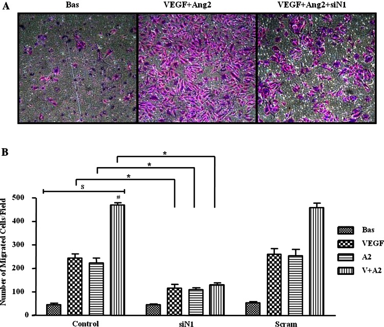
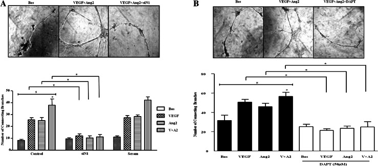
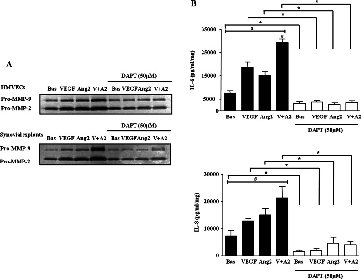
Methods: Notch-1 intracellular domain (Notch-1 IC), Notch-4 IC, Delta-like-ligand 4, Hes-related transcriptional repressors-1 and 2 (Hrt-1, Hrt-2) mRNA and/or protein expression was measured by Real-time PCR and/or western blot. VEGF/Ang2 induced EC function was assessed using transwell invasion chambers, matrigel tube formation assays and wound repair scratch assays±Notch-1 siRNA or an γ-secretase inhibitor N-(N-(3,5-Difluorophenacetyl-L-alanly))-S-phenylglycine-t-Butyl Ester (DAPT) in RA synovial explants or human microvascular EC. Interleukin (IL)-6 and IL-8 were measured by ELISA and MMP2 and 9 by gelatine zymography.

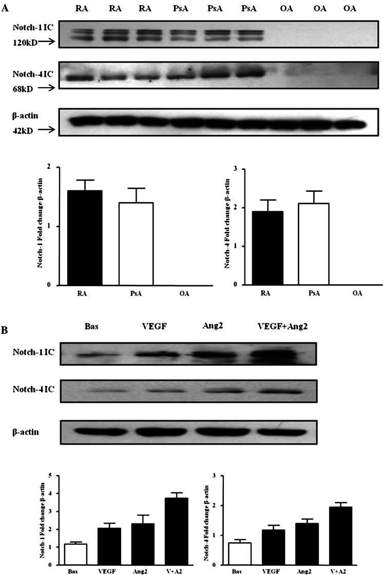
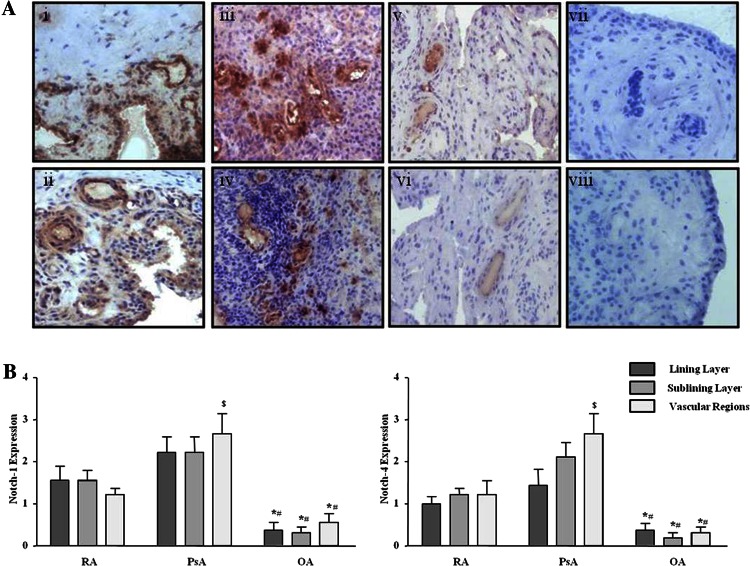
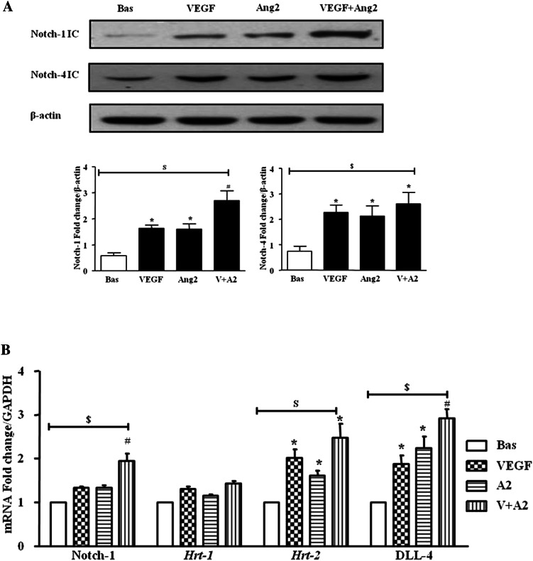
Results: Notch-1 IC and Notch-4 IC protein expressions were demonstrated in RA and psoriatic arthritis synovial biopsies, with minimal expression observed in Osteoarthritis (OA). VEGF and Ang2 induced Notch-1 IC/ Notch-4 IC protein expression in synovial explant cultures and human microvascular EC levels were further potentiated by VEGF/Ang2 stimulation in combination. Notch-1, Delta-like-ligand 4, and Hrt-2 mRNA expression were significantly induced by VEGF and Ang2 alone and in combination. Furthermore VEGF/Ang2-induced EC invasion, angiogenesis and migration were inhibited by Notch-1 siRNA or DAPT. Conditioned media from VEGF/Ang2 stimulated RA synovial explants induced EC tube formation, an effect that was inhibited by DAPT. Finally, DAPT significantly decreased VEGF/Ang2 induced IL-6, IL-8, MMP2 and 9 expressions in RA synovial explants.

Conclusions: Notch-1 mediates VEGF/Ang2-induced angiogenesis and EC invasion in inflammatory arthritis.

Keywords: Autoimmune Diseases; Inflammation; Rheumatoid Arthritis.


Figures

Figure 1
Figure 1
Vascular endothelial growth factor (VEGF) and angiopoietin 2 (Ang2) regulate Notch protein expression in synovial explant cultures. (A) Notch-1 intracellular domain (Notch-1 IC) and Notch-4 IC protein expression were analysed using Western blot in synovial tissues biopsies obtained from RA (n=7), psoriatic arthritis (n=7) and OA (n=6) patients. β-actin was used as loading control. (B) Notch-1 IC and Notch-4 IC protein expression was examined in ex vivo RA synovial explants cultures following stimulation with VEGF (20 ng/ml) and Ang2 (250 ng/ml) alone or in combination for 24 h. β-actin was used as loading control. Representative blot of n=3 experiments performed in triplicate.
Figure 2
Figure 2
Notch-1 and Notch-4 localised expression in RA, psoriatic arthritis (PsA) and OA patients. (A) Representative photomicrographs showing Notch-1 and Notch-4 localised expression in synovial tissue from RA (n=10), PsA (n=10) and OA (n=9) patients. Notch-1 and Notch-4 expression are shown in RA tissue (i, ii), PsA (iii, iv), OA (v, vi) respectively. Negative staining for IgG control (vii, viii). (B) Quantification of Notch-1 and Notch-4 in lining, sublining and perivascular regions of RA, PsA and OA tissue. *p<0.05 OA significantly different from RA, #p<0.05 OA significantly different from PsA, $p<0.05 PsA significantly different from RA. Original magnification ×10.
Figure 3
Figure 3
Vascular endothelial growth factor (VEGF) and angiopoietin 2 (Ang2) regulate the Notch signalling pathway in human microvascular endothelial cell (HMVEC). (A) Notch-1 intracellular domain (Notch-1 IC) and Notch-4 IC protein expression was examined in HMVEC following stimulation with VEGF (20 ng/ml) and Ang2 (250 ng/ml) alone or in combination for 24 h by Western blot. β-actin was used as loading control. Representative blot of n=4 experiments. (B) Notch-1, Hrt-1. Hrt-2 and DLL-4 mRNA expression was quantified in HMVEC following stimulation with VEGF (20 ng/ml) and Ang2 (250 ng/ml) alone or in combination for 24 h and normalised to Glyceraldehyde 3-phosphate dehydrogenase (GAPDH). Data is representative of three independent experiments. Values are expressed as mean±SEM of n=3 experiments. $p<0.05 significantly different for comparison of multiple parameters by ANOVA, *p<0.05 significantly different from basal, #p<0.05 significantly different from VEGF.
Figure 4
Figure 4
Vascular endothelial growth factor (VEGF) and angiopoietin 2 (Ang2)-induced cell invasion is dependent on Notch signalling. (A) Representative photomicrograph shows human microvascular endothelial cell (HMVEC) invasion under basal conditions (left panel), following VEGF/Ang2 stimulation (middle panel) and in the presence of Notch-1 siRNA (siN1, right panel). At 24 h invading cells attached to lower membrane were fixed (1% glutaraldehyde) and stained (0.1% crystal violet) (hpf, magnification × 40). (B) Representative graphs quantifying non-transfected (control), transfected (Notch-1 or Scramble siRNA), HMVEC invasion following stimulation with VEGF (20 ng/ml), and Ang2 (250 ng/ml) alone or in combination for 16 h. Data are expressed as mean±SEM of replicate experiments (n=4). $p<0.05 significantly different for comparison of multiple parameters by ANOVA, #p<0.05 significantly different from VEGF, *p<0.05 significantly different.
Figure 5
Figure 5
Notch signalling mediates vascular endothelial growth factor (VEGF) and angiopoietin 2 (Ang2)-induced cell tube formation. (A) Representative photomicrographs showing human microvascular endothelial cell (HMVEC) tube formation at baseline (left panel), following stimulation with VEGF and Ang2 (middle panel), and in the presence of Notch-1 siRNA transfection (right panel). Quantification of HMVEC tube formation following transfection with Notch-1 or Scramble siRNA following stimulation with VEGF (20 ng/ml) and Ang2 (250 ng/ml) alone or in combination. Data are expressed as mean±SEM of replicate experiments (n=4). (B) Representative photomicrograph shows HMVEC tube formation in basal conditioned media containing DMSO as vehicle control (left panel), conditioned media containing VEGF/Ang2 combination (middle panel), and in the presence of 50 µM N-(N-(3,5-Difluorophenacetyl-L-alanly))-S-phenylglycine-t-Butyl Ester (DAPT) (right panel). Connecting branches numbers were quantified following stimulation with VEGF (20 ng/ml) and Ang2 (250 ng/ml) alone or in combination. Data are expressed as mean±SEM of replicate experiments (n=4). $p<0.05 significantly different for comparison of multiple parameters by ANOVA or by Friedman analysis, #p<0.05 significantly different from VEGF, *p<0.05 significantly different.
Figure 6
Figure 6
N-(N-(3,5-Difluorophenacetyl-L-alanly))-S-phenylglycine-t-Butyl Ester (DAPT) inhibits vascular endothelial growth factor (VEGF)/angiopoietin 2 (Ang2) induced MMPs and cytokines activities. (A) Representative in-gel zymography of pro-MMP-9 and MMP-2 activities (inverted for clarity) in human microvascular endothelial cells (HMVEC) and synovial explants following stimulation with VEGF (20 ng/ml) and Ang2 (250 ng/ml) alone or in combination in the presence of DAPT (50 µM) or DMSO vehicle control. (B) Levels of IL-6 and IL-8 were assayed by ELISA in conditioned media with DAPT (50 µM). Values are expressed as mean±SEM of replicate experiments (n=5). $p<0.05 significantly different for comparison of multiple parameters by ANOVA or by Friedman analysis, #p<0.05 significantly different from VEGF, *p<0.05 significantly different.

References

    1. Akil M, Amos RS. ABC of rheumatology. Rheumatoid arthritis—I: clinical features and diagnosis. BMJ 1995;310:587–90 - PMC - PubMed
    1. Folkman J, D'Amore PA. Blood vessel formation: what is its molecular basis? Cell 1996;87:1153–5 - PubMed
    1. Koch AE. Angiogenesis as a target in rheumatoid arthritis. Ann Rheum Dis 2003;62(Suppl 2):ii60–7 - PMC - PubMed
    1. Reece RJ, Canete JD, Parsons WJ, et al. Distinct vascular patterns of early synovitis in psoriatic, reactive, and rheumatoid arthritis. Arthritis Rheum 1999;42:1481–4 - PubMed
    1. Fearon U, Griosios K, Fraser A, et al. Angiopoietins, growth factors, and vascular morphology in early arthritis. J Rheumatol 2003;30:260–8 - PubMed

Publication types

MeSH terms