. 2012 Dec 11;109(50):E3424-33.
doi: 10.1073/pnas.1203035109. Epub 2012 Nov 16.

Structure of the formin-interaction domain of the actin nucleation-promoting factor Bud6

Affiliations

Structure of the formin-interaction domain of the actin nucleation-promoting factor Bud6

Daqi Tu et al. Proc Natl Acad Sci U S A. .

Abstract

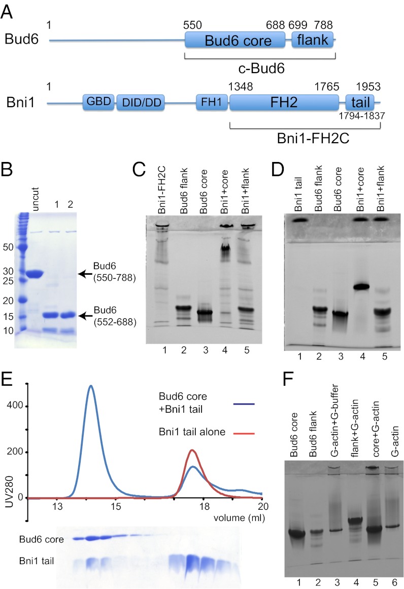
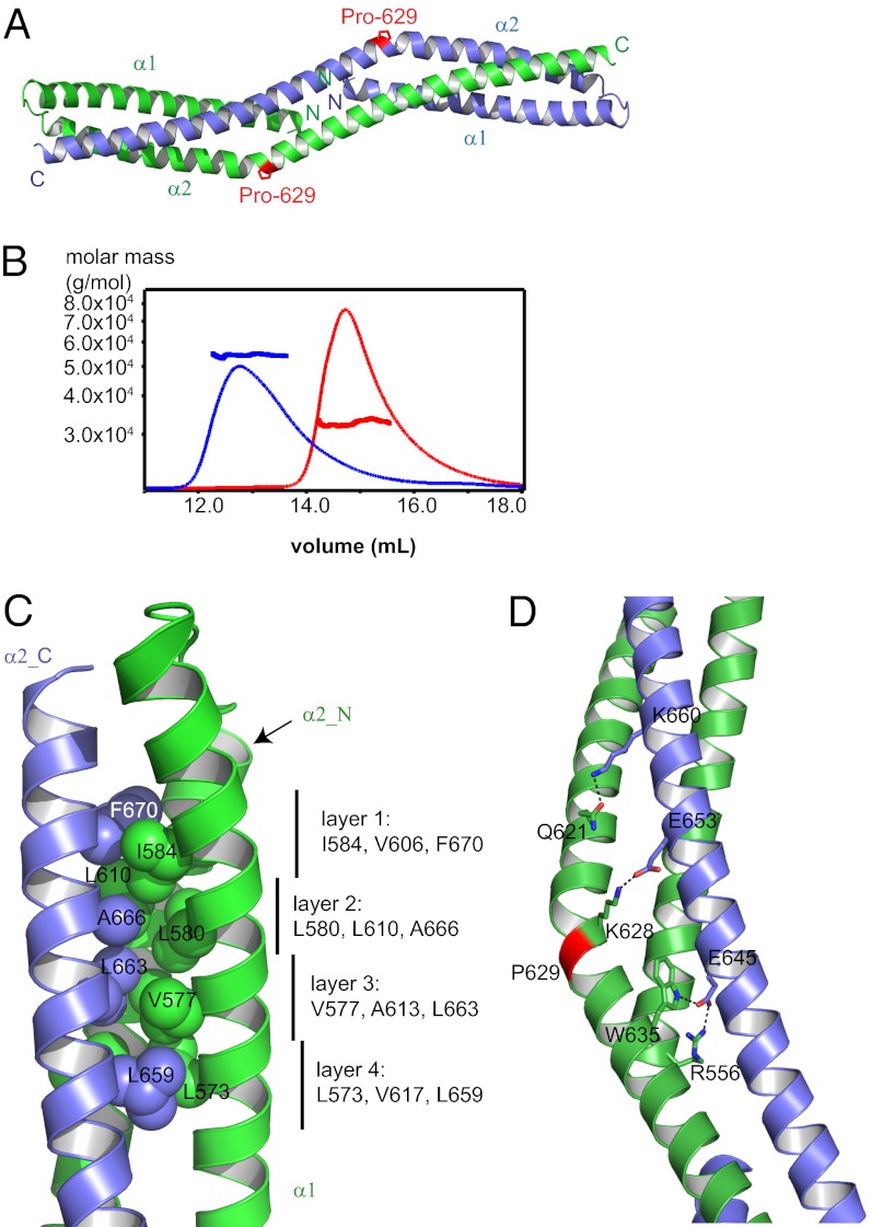
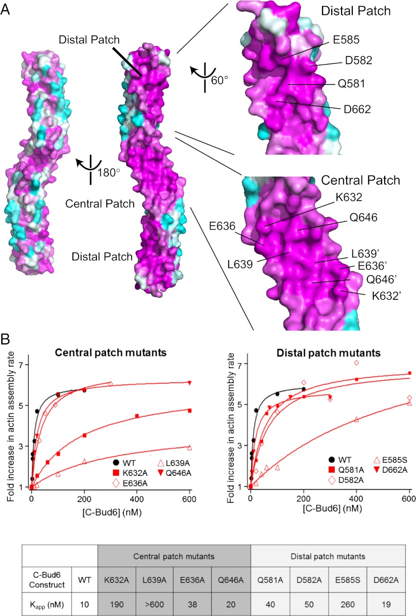
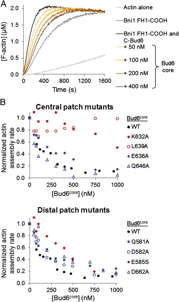
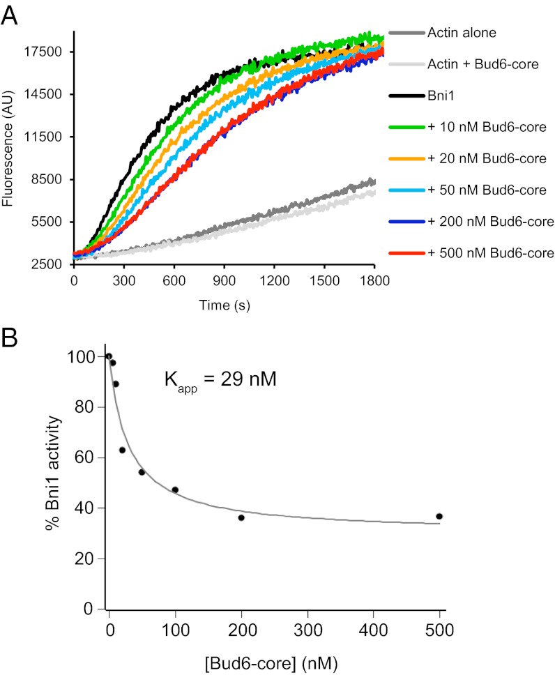
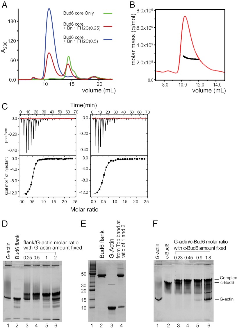
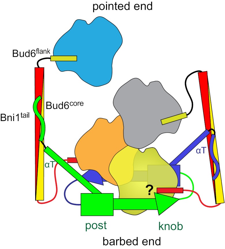
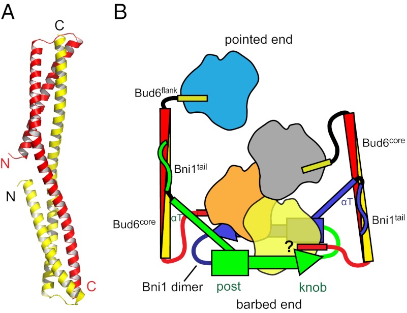
Formin proteins and their associated factors cooperate to assemble unbranched actin filaments in diverse cellular structures. The Saccharomyces cerevisiae formin Bni1 and its associated nucleation-promoting factor (NPF) Bud6 generate actin cables and mediate polarized cell growth. Bud6 binds to both the tail of the formin and G-actin, thereby recruiting monomeric actin to the formin to create a nucleation seed. Here, we structurally and functionally dissect the nucleation-promoting C-terminal region of Bud6 into a Bni1-binding "core" domain and a G-actin binding "flank" domain. The ∼2-Å resolution crystal structure of the Bud6 core domain reveals an elongated dimeric rod with a unique fold resembling a triple-helical coiled-coil. Binding and actin-assembly assays show that conserved residues on the surface of this domain mediate binding to Bni1 and are required for NPF activity. We find that the Bni1 dimer binds two Bud6 dimers and that the Bud6 flank binds a single G-actin molecule. These findings suggest a model in which a Bni1/Bud6 complex with a 2:4 subunit stoichiometry assembles a nucleation seed with Bud6 coordinating up to four actin subunits.


Conflict of interest statement

The authors declare no conflict of interest.

Figures

Fig. 1.
Fig. 1.
Dissection of c-Bud6 functional domains. (A) Domain structures of Bud6 and Bni1. The residue boundaries of the constructs studied here are highlighted. (B) Trypsin digest of c-Bud6. Aliquots of c-Bud6 alone (uncut) or c-Bud6 treated with trypsin for the indicated time (in hours) were analyzed by Coomassie-stained SDS/PAGE. (C) Native-PAGE analysis of Bni1/Bud6 complexes. Bni1-FH2C was mixed with either Bud6core or Bud6flank and resolved on an 8–25% gradient native gel. Note that Bni1-FH2C does not migrate as a single species but that it completely shifts the well-defined Bud6core band to a much slower migrating position. (D) Native-PAGE analysis of the Bni1tail and its interactions with Bud6core and Bud6flank. The proteins alone (lanes 1–3) or mixtures of an excess of Bni1tail with Bud6core (lane 4) or Bud6flank (lane 5) were resolved on 20% native gel. Note that Bni1tail completely shifts the Bud6core band. (E) SEC of the Bni1 tail region alone (red trace) or mixed in excess with the Bud6core protein (blue trace). SDS/PAGE analysis of the elution fractions from the Bni1/Bud6 mixture (Lower) shows that a fraction of the Bni1 tail protein elutes together with the Bud6core, indicating that the complex is stable on gel filtration. (F) Native-PAGE analysis of Bud6core, Bud6flank, and G-actin. The Bud6 proteins alone or mixed with G-actin in G-buffer, as indicated, were resolved on 20% native gel. Note that Bud6flank shifts the G-actin band to a more slowly migrating position, indicating formation of a complex.
Fig. 2.
Fig. 2.
Structure of Bud6core. (A) Overall structure of Bud6core dimer. Two chains are colored green and blue. The proline residue, Pro-629, that causes sharp bend of the second long helix is indicated by a stick representation of its side chain, in magenta. (B) SEC-MALS analysis of Bud6core and c-Bud6 reveals that both are dimers in solution. Purified Bud6core (residues 550–688) and c-Bud6 (residues 550–788) were analyzed on a Superdex 200 gel filtration column coupled to a MALS detector. The elution profile as measured by refractive index is shown for Bud6core as a thin red trace and for c-Bud6 as a thin black trace. The thicker horizontal traces indicate the measured molar mass, ∼32.7 kDa for Bud6core (expected molar mass for a Bud6core dimer is 32.7 kDa) and ∼54.4 kDa for c-Bud6 (theoretical molar mass is 55.6 kDa for a c-Bud6 dimer). (C) Intersubunit hydrophobic interactions of the Bud6core. Chain A is shown in green, and chain B is shown in blue. Designations of α1, α2_N, and α2_C are the same as in Fig. S4B. Side chains are shown in sphere presentation with the Van der Waals radius. (D) Number of polar interactions also stabilizes the dimer; hydrogen bonds are indicated by dashed lines.
Fig. 3.
Fig. 3.
Identification of conserved functional surface residues on Bud6core. (A) Surface conservation of Bud6core. Each residue is labeled with a color ranging from the most conserved (magenta) to the most variable (cyan) as analyzed with ConSurf (37). The eight residues that were mutated for biochemical analysis are indicated on the surface in the zoomed-in view. The two identical central patches are contiguous; residues contributing to these patches are distinguished by the use of prime symbols in their labels. (B) Concentration-dependent effects of WT and mutant c-Bud6 polypeptides on Bni1-mediated actin assembly. Monomeric actin (2 μM, 2.5% pyrene-labeled) was polymerized in the presence of 10 nM Bni1 (FH1-FH2-C) and variable concentrations of c-Bud6 as indicated. Each data point is an average of at least two trials in which the maximum rate of actin assembly over the course of the reaction was determined. All values were normalized to the rate of actin assembly occurring in the presence of 10 nM Bni1 alone. The Kapp value for each mutant was calculated by determining the concentration of mutant c-Bud6 required to increase the rate of Bni1-mediated assembly to half of the maximal rate observed for WT c-Bud6.
Fig. 4.
Fig. 4.
Competition of Bud6core and c-Bud6 polypeptides for binding to Bni1 (FH1-FH2-C). (A) Monomeric actin (2 μM) was assembled in the presence of 10 nM Bni1 (FH1-FH2-C), 100 nM WT c-Bud6, and indicated concentrations of WT Bud6core. (B) Reactions were performed as in A, except using the indicated mutant Bud6core polypeptides. Each data point represents a single trial, where the slope of the raw curve was measured at 50% polymerization. Red datasets indicate mutations that impair the NPF activity of c-Bud6 (Fig. 3B); blue datasets indicate mutations in c-Bud6 that were pseudo-WT for stimulating Bni1.
Fig. 5.
Fig. 5.
Bud6core domain inhibits Bni1-mediated actin assembly. (A) Monomeric actin (2 μM, 5% pyrene-labeled) was assembled in the presence of 10 nM C-Bni1 (FH1-FH2-C) and a range of concentrations of Bud6core domain. AU, arbitrary units; These are the raw fluorescence readings. (B) Concentration-dependent effects of Bud6core domain on the rate of C-Bni1–mediated actin assembly. The percentage of activity was determined from the slopes of the raw curves as in A. The slope for C-Bni1 in the absence of Bud6 was defined as 100% activity. Each data point is an average of two independent trials.
Fig. 6.
Fig. 6.
Stoichiometry of Bud6 interaction with Bni1 and G-actin. (A) Analysis of Bni1-FH2C/Bud6core mixtures by SEC. Bud6core alone, or with addition of a 0.25 or 0.5 molar ratio of Bni1-FH2C as indicated, was analyzed by SEC on a Superdex 200 column. Note that a 0.5 ratio of Bni1-FH2C was sufficient to ablate the free Bud6core peak (blue trace). Quantitation of Bud6core and Bni1-FH2C in the peak eluting at ∼11 mL by densitometry of the elution fractions resolved by SDS/PAGE confirmed a 2:1 ratio of Bud6core to Bni1-FH2C. (B) SEC-MALS analysis of c-Bud6/Bni1-FH2C indicated a molar mass of ∼240 kDa. Molar mass (black) and refractive index (red) are plotted vs. elution volume from a Superdex 200 size-exclusion column. A 2:1 complex of c-Bud6/Bni1 dimers has an expected mass of ∼250 kDa. (C) Analysis of Bud6 binding to Bni1tail by ITC. (Left) Titration of Bud6core with Bni1tail yielded a stoichiometry of n = 0.48 and Kd = 0.75 μM. (Right) Titration of c-Bud6 with Bni1tail produced similar results (n = 0.49 and Kd = 1.1 μM). (D) Native-PAGE analysis indicating that Bud6flank forms a 1:1 complex with G-actin. The amount of G-actin was fixed, and the amount of Bud6flank was gradually increased by the molar ratio indicated. (E) SDS/PAGE analysis of bands from a Bud6flank/G-actin native-PAGE gel shift assay. The upper bands from Bud6flank/G-actin molar ratios of 1 and 2 in D were excised, combined, and resolved on a 20% SDS/PAGE denaturing gel. (F) Native-PAGE analysis indicates that c-Bud6 forms a 1:1 complex with G-actin. A fixed amount of c-Bud6 was combined with an increasing molar ratio of G-actin as indicated and analyzed on 20% native gel. All protein concentrations were determined by amino acid analysis.
Fig. 7.
Fig. 7.
Structurally informed model for collaboration between Bud6 and Bni1 in actin filament nucleation. The figure is drawn based on the structural and binding data presented here, as well as structures of Bni1 and its complex with actin (12, 13). The two sides of the formin dimer are shown in green and purple; actin subunits are colored yellow, orange, gray, and cyan; and the Bud6 dimer is shown in yellow and red. The Bni1 FH2 dimer binds two Bud6 dimers, which, in turn, bind a total of four actin subunits to assemble a nucleation seed with Bni1. It is important to note that the number of actin subunits required for productive nucleation by Bni1 and Bud6 remains to be determined and the depiction of which actin subunits are coordinated by the Bud6 flank domains is hypothetical. It is possible that the Bud6flank dissociates from actin on formation of a stable nucleus, because c-Bud6 binds to G-actin but not F-actin. Additionally, it is unclear whether Bud6 and Bni1 can simultaneously engage the same actin subunit, because the Bud6flank binding site on actin remains to be elucidated.
Fig. P1.
Fig. P1.
Structure of the Bud6core domain and a model for actin filament nucleation by the Bni1/Bud6 complex. (A) Ribbon diagram of the Bud6core domain, with the N- and C-terminal ends of the fragment labeled and with one chain of the dimer colored red and the other yellow. (B) Working model for actin nucleation on Bni1, derived from the work described here and from structures of the Bni1 FH2 domain alone and with actin (5). The two sides of the formin dimer are shown in green and blue, and the actin subunits are shown in yellow, orange, gray, and blue. The Bni1 FH2 dimer binds two Bud6 dimers, and Bud6, in turn, may bring a total of four actin subunits into the nascent filament. After the nucleus is formed, Bud6 may remain associated or dissociate from Bni1 as actin elongation proceeds, with the formin processively tracking the barbed end of the growing filament. As indicated by the question mark, it is unclear whether Bud6 and Bni1 can simultaneously engage the same actin subunit.

Similar articles

Cited by

References

    1. Chesarone MA, Goode BL. Actin nucleation and elongation factors: Mechanisms and interplay. Curr Opin Cell Biol. 2009;21(1):28–37. - PMC - PubMed
    1. Dominguez R. Actin filament nucleation and elongation factors—Structure-function relationships. Crit Rev Biochem Mol Biol. 2009;44(6):351–366. - PMC - PubMed
    1. Campellone KG, Welch MD. A nucleator arms race: Cellular control of actin assembly. Nat Rev Mol Cell Biol. 2010;11(4):237–251. - PMC - PubMed
    1. Goode BL, Eck MJ. Mechanism and function of formins in the control of actin assembly. Annu Rev Biochem. 2007;76:593–627. - PubMed
    1. Sept D, McCammon JA. Thermodynamics and kinetics of actin filament nucleation. Biophys J. 2001;81(2):667–674. - PMC - PubMed

Publication types

MeSH terms

Associated data

LinkOut - more resources