. 2012 Nov 16;338(6109):949-53.
doi: 10.1126/science.1227157.

Pathological α-synuclein transmission initiates Parkinson-like neurodegeneration in nontransgenic mice

Affiliations

Pathological α-synuclein transmission initiates Parkinson-like neurodegeneration in nontransgenic mice

Kelvin C Luk et al. Science. .

Abstract

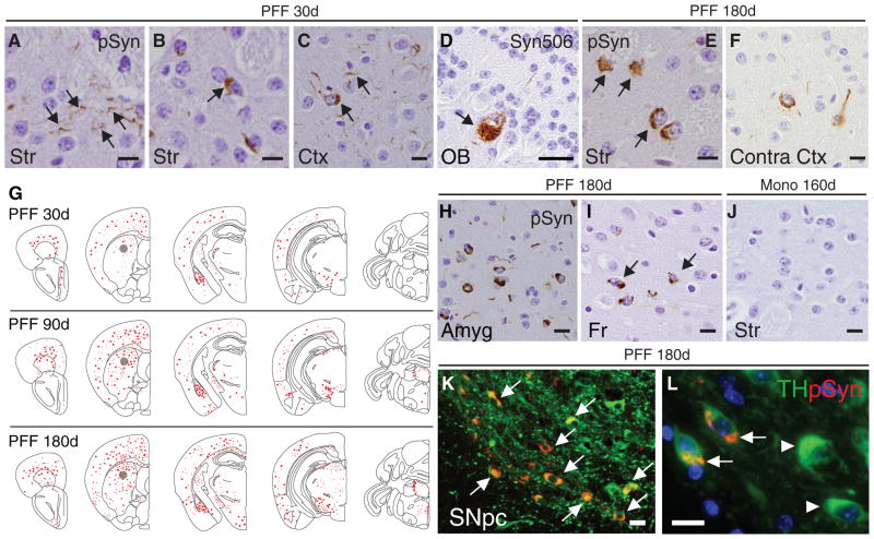
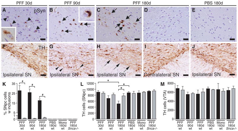
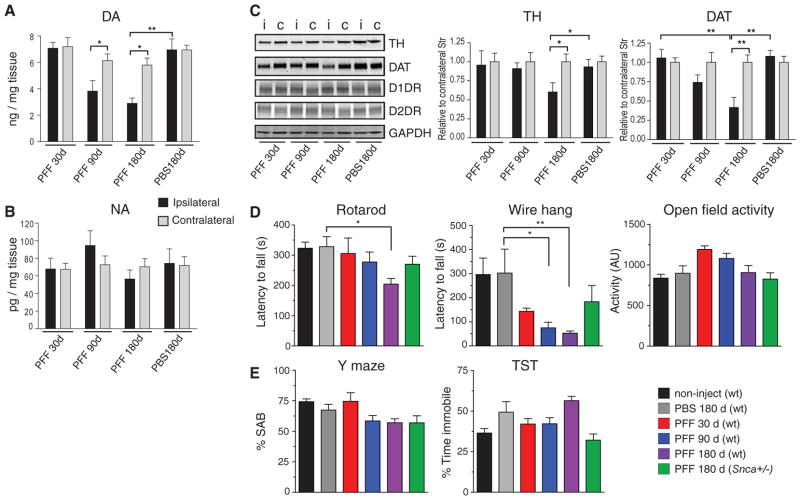
Parkinson's disease is characterized by abundant α-synuclein (α-Syn) neuronal inclusions, known as Lewy bodies and Lewy neurites, and the massive loss of midbrain dopamine neurons. However, a cause-and-effect relationship between Lewy inclusion formation and neurodegeneration remains unclear. Here, we found that in wild-type nontransgenic mice, a single intrastriatal inoculation of synthetic α-Syn fibrils led to the cell-to-cell transmission of pathologic α-Syn and Parkinson's-like Lewy pathology in anatomically interconnected regions. Lewy pathology accumulation resulted in progressive loss of dopamine neurons in the substantia nigra pars compacta, but not in the adjacent ventral tegmental area, and was accompanied by reduced dopamine levels culminating in motor deficits. This recapitulation of a neurodegenerative cascade thus establishes a mechanistic link between transmission of pathologic α-Syn and the cardinal features of Parkinson's disease.


Figures

Fig. 1
Fig. 1. Intrastriatal inoculation of synthetic mouse α-Syn PFFs seed the aggregation of endogenous mouse Syn in wt mice
Pathology in brains of C57BL6/C3H F1 mice following a single unilateral injection of mouse α-Syn PFFs into the dorsal striatum. (A–D) Accumulation of α-Syn in neuritic processes or as pale cytoplasmic inclusions in striatum (Str) and neocortex (Ctx), and olfactory mitral neurons (OB) ipsilateral to the injection at 30d. Black arrows highlight pathology revealed by immunostaining using anti-pSyn (A–C) or Syn506 (D). (E,F) LB-like inclusions in striatum and contralateral neocortex at 180d post-injection with PFFs. (G) CNS distribution of pSyn accumulations of mice that received a single inoculation of PFFs in the dorsal striatum (indicated by grey circles). Representative maps of LB/LN-like pathology (red dots and stipples, respectively) in the PFF-injected hemisphere are shown for mice sacrificed at 30d, 90d, or 180d post-injection. (H) α-Syn pathology in amygdala (Amyg), and (I) in frontal cortex (Fr). (J) pSyn staining in ipsilateral striatum 160d post-injection with monomeric recombinant α-Syn. (K) Double-immunostaining for pSyn (red) and TH (green) in a PFF-injected animal sacrificed at 180d showing LB-like α-Syn pathology in ipsilateral SNpc. (L) High magnification revealing colocalization of pSyn inclusions to DA neurons (white arrows) and reduced TH immunoreactivity compared to unaffected DA neurons (white arrowheads). Images are representative of 3–7 animals examined per group (see Table S1). Scale bars: 10 μm (A–C, E–F, H–L); 25 μm (D).
Fig. 2
Fig. 2. Seeded α-Syn pathology leads to progressive DA system degeneration
(A–D) pSyn immunostaining in SNpc of mice sacrificed at 30d, 90d, or 180d following striatal PFF injection. (A) Diffuse perinuclear pSyn inclusions (black arrowheads) at 30d post-injection. (B,C) Dense LB-like inclusions (black arrows) at 90d and 180d post-injection. Absence of pSyn pathology in SNpc contralateral to the PFF injection site (D) and in ipsilateral SNpc of PBS-injected mouse (E) at 180d. (F–I) TH-immunostaining of SNpc at 30d, 90d and 180d following inoculation with Syn PFFs. Arrowheads in (G) indicate neurons with reduced TH staining in the ipsilateral SNpc at 90d post-injection. (H,I) Ventral SNpc, ipsilateral and contralateral to the site of PFF injection at 180d. Arrows point to DA neuron loss. (J) PBS-injected control at 180d post-injection. (K) Percentage of SNpc TH-neurons containing pSyn-immunoreactive inclusions for each treatment group. Data for treated ipsilateral (black) and contralateral (grey) hemispheres are shown. #p<0.001 paired t-test (N = 3–5 animals per group). (L,M) Quantification of TH-immunoreactive neurons in the SNpc and VTA of mice after intrastriatal PFF-, monomer- or PBS-injection. Data represent mean number of cells per region +/− SEM (N = 3–4 animals per group). *P<0.05 one-way ANOVA. Scale bars: 25 μm (A–D);50 μm (E–J).
Fig. 3
Fig. 3. Decreased striatal DA and motor deficits in mouse α-Syn PFF-injected wt mice
DA (A) and NA (B) concentrations in dorsal striata of PFF-injected wt mice and PBS-treated controls measured at the indicated time points. Mean +/− SD are shown (N = 3–6 animals per group. *P<0.01; **P<0.001 one-way ANOVA. (C) Immunoblot analysis of striata from PFF- and PBS-treated animals, using antibodies against TH, DAT, D1 dopamine receptor (D1DR), and D2 dopamine receptor (D2DR). GAPDH is shown as a loading control. Mean intensity values are shown for TH and DAT (N = 3–4 striata per marker). Black and grey bars denote ipsilateral and contralateral regions, respectively. *P<0.05; **P<0.001, one-way ANOVA. (D,E) Behavioral assessment of wt mice 30d, 90d, or 180d (N = 6, 9, 17, and 6, respectively) following a single unilateral inoculation of α-Syn PFFs into the striatum. PFF-injected Snca+/− mice (N = 4), as well as age-matched non-injected (N = 20) and PBS-injected (N = 8) wt animals are also shown. Results of animals on the rotarod test (D, left panel) and wire hang test (D, middle panel) show progressive deficits in PFF-injected but not control mice. (E) Performance on the Y-maze and tail suspension test. Data are mean values +/− SD. Differences were established using one-way ANOVA (P=0.0012) with Tukey post-hoc test (*P<0.05; **P<0.01).

Comment in

Similar articles

Cited by

References

    1. Fearnley JM, Lees AJ. Ageing and Parkinson’s disease: substantia nigra regional selectivity. Brain. 1991;114:2283. - PubMed
    1. Simon-Sanchez J, et al. Genome-wide association study reveals genetic risk underlying Parkinson’s disease. Nat Genet. 2009;41:1308. - PMC - PubMed
    1. Singleton AB, et al. alpha-synuclein locus triplication causes Parkinson’s disease. Science. 2003;302:841. - PubMed
    1. Poulopoulos M, Levy OA, Alcalay RN. The neuropathology of genetic Parkinson’s disease. Mov Disord. 2012;27:831. - PMC - PubMed
    1. Spillantini MG, et al. alpha-synuclein in Lewy bodies. Nature. 1997;388:839. - PubMed

Publication types

MeSH terms