. 2013 Jan;33(1):e1-e10.
doi: 10.1161/ATVBAHA.112.300399. Epub 2012 Nov 15.

Loss of CDKN2B promotes p53-dependent smooth muscle cell apoptosis and aneurysm formation

Affiliations

Loss of CDKN2B promotes p53-dependent smooth muscle cell apoptosis and aneurysm formation

Nicholas J Leeper et al. Arterioscler Thromb Vasc Biol. 2013 Jan.

Abstract

Objective: Genomewide association studies have implicated allelic variation at 9p21.3 in multiple forms of vascular disease, including atherosclerotic coronary heart disease and abdominal aortic aneurysm. As for other genes at 9p21.3, human expression quantitative trait locus studies have associated expression of the tumor suppressor gene CDKN2B with the risk haplotype, but its potential role in vascular pathobiology remains unclear.

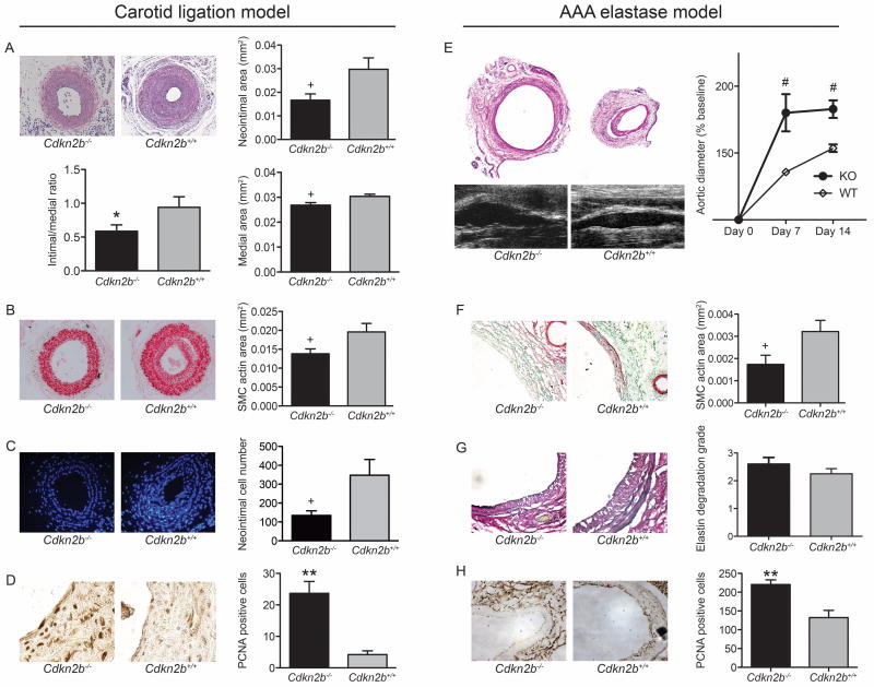
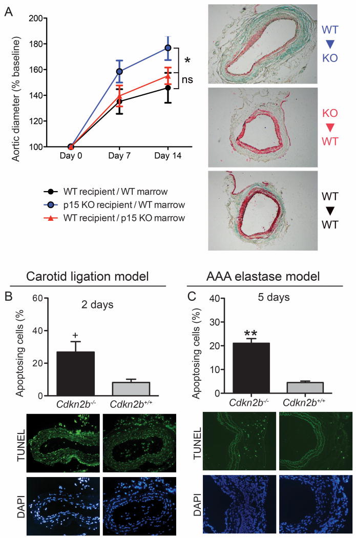
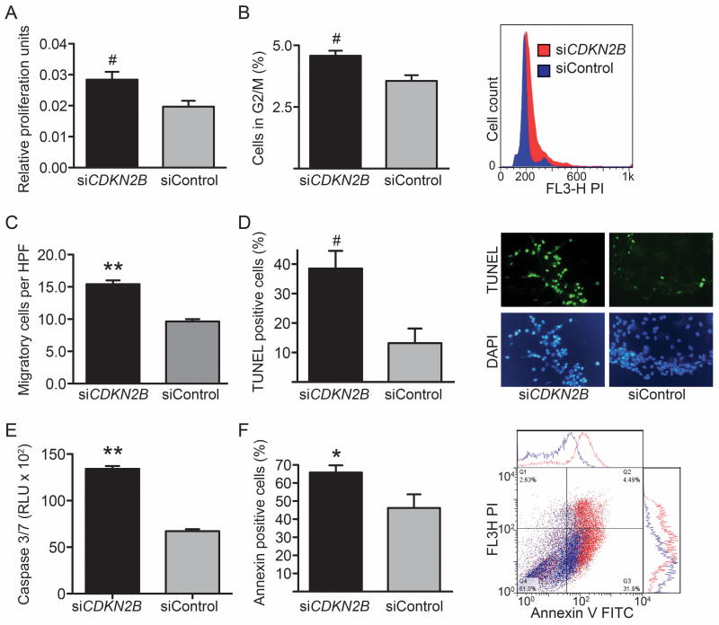
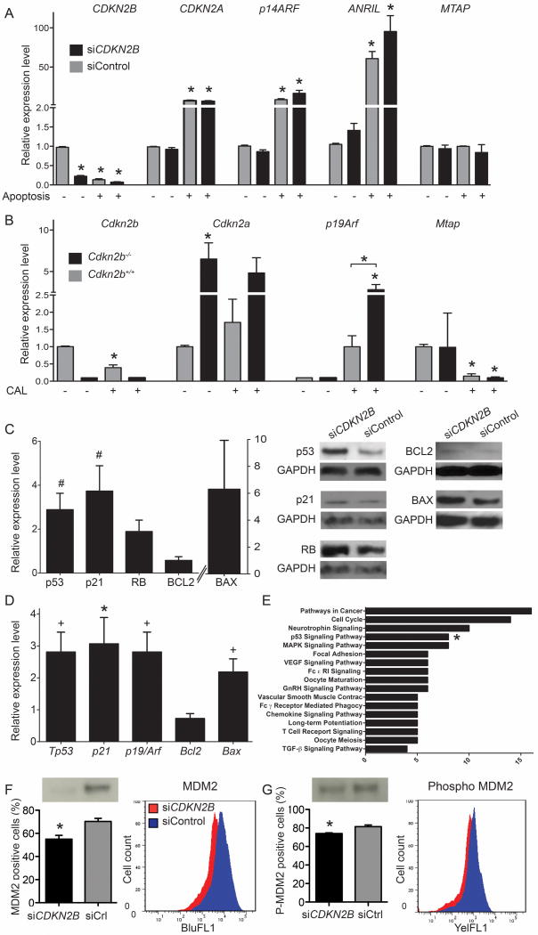
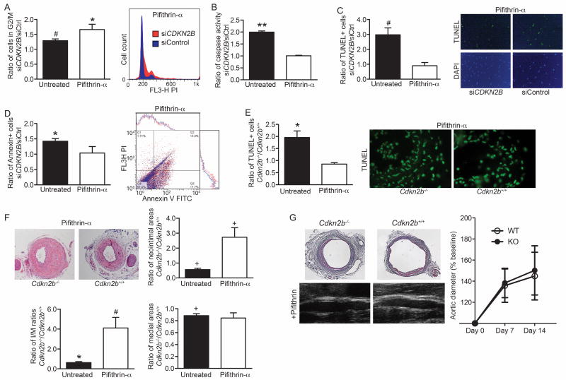
Methods and results: Here we used vascular injury models and found that Cdkn2b knockout mice displayed the expected increase in proliferation after injury, but developed reduced neointimal lesions and larger aortic aneurysms. In situ and in vitro studies suggested that these effects were attributable to increased smooth muscle cell apoptosis. Adoptive bone marrow transplant studies confirmed that the observed effects of Cdkn2b were mediated through intrinsic vascular cells and were not dependent on bone marrow-derived inflammatory cells. Mechanistic studies suggested that the observed increase in apoptosis was attributable to a reduction in MDM2 and an increase in p53 signaling, possibly due in part to compensation by other genes at the 9p21.3 locus. Dual inhibition of both Cdkn2b and p53 led to a reversal of the vascular phenotype in each model.

Conclusions: These results suggest that reduced CDKN2B expression and increased smooth muscle cell apoptosis may be one mechanism underlying the 9p21.3 association with aneurysmal disease.


Figures

Figure 1
Figure 1
Cdkn2b regulates murine vascular disease. (A) Cdkn2b-/- mice display alterations in vascular remodeling in the carotid ligation model 28 days after vascular injury. Compared to Cdkn2b+/+ mice (right, n=15), Cdkn2b-/- mice (left, n=19) have smaller neointimal areas (upper right), medial areas (lower right) and intimal-to-medial ratios (lower left). (B) Cdkn2b-/- vessels have less SMC content as measured by SMC α-actin positive area, and (C) fewer total vascular cells as assessed by number of DAPI positive nuclei. (D) Cdkn2b-/- mice displayed higher rates of cell division as assessed by PCNA stain. (E) Cdkn2b-/- mice (n= 9) also develop significantly larger aortic aneurysms in the elastase infusion model than Cdkn2b+/+ mice (n=8), with (F) associated decrease in SMC number, and no significant difference in (G) elastin degradation score. (H) As in the CAL model, there was a significant increase in cell proliferation in aortas of elastase model Cdkn2b-/- mice. * = P < 0.05; + = P < 0.03; # = P < 0.01; ** = P < 0.001.
Figure 2
Figure 2
Cdkn2b regulates apoptosis in vivo and potentiates disease through intrinsic vessel wall cells. (A) Bone marrow transplant studies revealed that Cdkn2b's vascular effects are not mediated through bone marrow-derived cells, with enhanced aneurysms only observed in the Cdkn2b-/-Recipient/WTDonor group compared to the WTRecipient/Cdkn2b-/-Donor and WTRecipient/WTDonor groups (n= 15 in each group, P<0.05). Similarly, a significant reduction in SMC α-actin content was only observed in the Cdkn2b-/-Recipient group. (B) Cdkn2b-/- mice (n= 8) displayed markedly elevated rates of vascular apoptosis relative to Cdkn2b+/+ mice (n= 8) two days after ligation (3.32 fold increase, P<0.03), as assessed by percentage of TUNEL-positive cells in the remodeling carotid artery. These differences were no longer observed four weeks after vascular injury. (C) Cdkn2b-/- mice (n=9) displayed increased aortic apoptosis relative to Cdkn2b+/+mice (n=5) five days after elastase infusion (4.64 fold increase, P<0.0001) in the AAA model.
Figure 3
Figure 3
CDKN2B regulates SMC functions in cultured human vascular smooth muscle cells. HCASMC deficient in CDKN2B (siCDKN2B) have higher rates of proliferation than control cells (siControl) as assessed by (A) MTT assay, and (B) percentage of cells in G2/M phase by FACS analysis. (C) siCDKN2B treated cells have higher rates of migration in the Boyden chamber assay than siControl cells. siCDKN2B treated cells have significantly higher rates of programmed cell death, as assessed by (D) TUNEL staining in chamber slides, (E) Caspase 3/7 activity, and (F) Annexin V positivity by FACS analysis, confirming the in vivo findings.
Figure 4
Figure 4
Compensation occurs at the 9p21 locus in response to stress and loss of CDKN2B promotes the activity of apoptotic factors, including MDM2 and p53. (A) In response to an apoptotic stimulus, control-transfected HCASMC (grey bars) display reduced expression of CDKN2B and increased expression of CDKN2A, ARF and ANRIL. A similar pattern is observed in CDKN2B-deficient cells (black bars). All comparisons are made to basal, control-transfected cells (*=P<0.05). (B) A similar pattern of compensation is observed in carotid tissue in vivo in wild type mice (n=7, grey bars) after carotid injury. Comparing the relative response to stress in Cdkn2b-/- mice (n=7, black bars) reveals that the change in Arf is accentuated compared to wild type animals (P<0.05). (C) Western blot analysis of apoptosing CDKN2B-deficient HCASMC also revealed a significant upregulation of p53 and the downstream p21 protein relative to control siRNA transfected cells. A trend towards increased expression of the pro-apoptotic protein BAX and reduced expression of the anti-apoptotic protein BCL-2 was also observed. RB expression was not significantly changed by knockdown of CDKN2B. Quantitative densitometric data is shown in graph format on the left, representative blots with GAPDH loading controls on the right. (D) mRNA expression analysis of mouse carotid tissue four days after carotid ligation confirm the in vitro findings, with significant upregulation of Trp53, p21, Arf and Bax observed in the vessels of Cdkn2b-/- mice (n= 7) relative to Cdkn2b+/+ mice (n= 7). (C) Phospho-antibody protein microarray studies and KEGG analysis in HCASMC confirm the central role of the p53 apoptotic pathway in CDKN2B signaling. Among the differentially regulated genes annotated under the p53 signaling pathway (asterisks), MDM2 was the most significantly altered. Flow cytometry assays and Western blot analyses confirmed the protein array data and reveal that apoptotic CDKN2B-deficient HCASMC express significantly less total MDM2 than apoptotic control transfected cells (D, P<0.01) as well as phosphorylated MDM2 (E, P<0.01).
Figure 5
Figure 5
CDKN2B's effects on remodeling and apoptosis are dependent on p53. In vitro, inhibition of both CDKN2B and p53 eliminated the difference in apoptosis while enhancing the difference in proliferation. (A) Inhibition of p53 with pifithrin-α accentuated the ratio of cell proliferation in CDKN2B deficient HCASMC relative to control transfected cells (siCDKN2B/siCtrl), compared to the baseline ratio. In contrast, treatment with pifithrin-α eliminated the difference in staurosporine-induced apoptosis that was noted at baseline, as assessed by (B) caspase 3/7 activity, (C) TUNEL positivity, and (D) annexin-V positivity by FACS analysis. In vivo, p53 inhibition also neutralized the difference in apoptosis between Cdkn2b-/- (n= 6) and Cdkn2b+/+ mice (n= 7), and led to a reversal of the remodeling phenotype compared to baseline. (E) One week after carotid ligation, the ratio of TUNEL positive cells between theCdkn2b-/- and Cdkn2b+/+ genotypes (Cdkn2b-/-/Cdkn2b+/+) had normalized in mice treated with intraperitoneal pifithrin-α, compared to baseline where the knockout mice had nearly double the rate of apoptosis (n= 5 for each). (F) Four weeks after ligation, Cdkn2b-/- mice treated with intraperitoneal pifithrin-α had larger neointimal areas and I/M ratios than wild type mice also treated with the p53 inhibitor, showing a reversal of phenotype after inhibiting the p53 pathway. (G) Similarly, aneurysm expansion was no longer different between genotypes after pifithrin-α treatment, (P > 0.7) at each timepoint (n=6 vs. 5).

Similar articles

  • CDKN2B Regulates TGFβ Signaling and Smooth Muscle Cell Investment of Hypoxic Neovessels.
    Nanda V, Downing KP, Ye J, Xiao S, Kojima Y, Spin JM, DiRenzo D, Nead KT, Connolly AJ, Dandona S, Perisic L, Hedin U, Maegdefessel L, Dalman J, Guo L, Zhao X, Kolodgie FD, Virmani R, Davis HR Jr, Leeper NJ. Nanda V, et al. Circ Res. 2016 Jan 22;118(2):230-40. doi: 10.1161/CIRCRESAHA.115.307906. Epub 2015 Nov 23. Circ Res. 2016. PMID: 26596284 Free PMC article.
  • MFAP4 Promotes Vascular Smooth Muscle Migration, Proliferation and Accelerates Neointima Formation.
    Schlosser A, Pilecki B, Hemstra LE, Kejling K, Kristmannsdottir GB, Wulf-Johansson H, Moeller JB, Füchtbauer EM, Nielsen O, Kirketerp-Møller K, Dubey LK, Hansen PB, Stubbe J, Wrede C, Hegermann J, Ochs M, Rathkolb B, Schrewe A, Bekeredjian R, Wolf E, Gailus-Durner V, Fuchs H, Hrabě de Angelis M, Lindholt JS, Holmskov U, Sorensen GL. Schlosser A, et al. Arterioscler Thromb Vasc Biol. 2016 Jan;36(1):122-33. doi: 10.1161/ATVBAHA.115.306672. Epub 2015 Nov 12. Arterioscler Thromb Vasc Biol. 2016. PMID: 26564819
  • H19 Induces Abdominal Aortic Aneurysm Development and Progression.
    Li DY, Busch A, Jin H, Chernogubova E, Pelisek J, Karlsson J, Sennblad B, Liu S, Lao S, Hofmann P, Bäcklund A, Eken SM, Roy J, Eriksson P, Dacken B, Ramanujam D, Dueck A, Engelhardt S, Boon RA, Eckstein HH, Spin JM, Tsao PS, Maegdefessel L. Li DY, et al. Circulation. 2018 Oct 9;138(15):1551-1568. doi: 10.1161/CIRCULATIONAHA.117.032184. Circulation. 2018. PMID: 29669788 Free PMC article.
  • Genetics of abdominal aortic aneurysm.
    Golledge J, Kuivaniemi H. Golledge J, et al. Curr Opin Cardiol. 2013 May;28(3):290-6. doi: 10.1097/HCO.0b013e32835f0d55. Curr Opin Cardiol. 2013. PMID: 23478885 Review.
  • The p53 pathway in vasculature revisited: A therapeutic target for pathological vascular remodeling?
    Cui X, Pan G, Chen Y, Guo X, Liu T, Zhang J, Yang X, Cheng M, Gao H, Jiang F. Cui X, et al. Pharmacol Res. 2021 Jul;169:105683. doi: 10.1016/j.phrs.2021.105683. Epub 2021 May 18. Pharmacol Res. 2021. PMID: 34019981 Review.

Cited by

References

    1. Lloyd-Jones DM, Nam BH, D'Agostino RB, Sr, Levy D, Murabito JM, Wang TJ, Wilson PW, O'Donnell CJ. Parental cardiovascular disease as a risk factor for cardiovascular disease in middle-aged adults: A prospective study of parents and offspring. JAMA. 2004;291:2204–2211. - PubMed
    1. Marenberg ME, Risch N, Berkman LF, Floderus B, de Faire U. Genetic susceptibility to death from coronary heart disease in a study of twins. N Engl J Med. 1994;330:1041–1046. - PubMed
    1. Musunuru K, Kathiresan S. Genetics of coronary artery disease. Annu Rev Genomics Hum Genet. 2010;11:91–108. - PubMed
    1. Schunkert H, Konig IR, Kathiresan S, et al. Large-scale association analysis identifies 13 new susceptibility loci for coronary artery disease. Nat Genet. 2011;43:333–338. - PMC - PubMed
    1. Helgadottir A, Thorleifsson G, Manolescu A, et al. A common variant on chromosome 9p21 affects the risk of myocardial infarction. Science. 2007;316:1491–1493. - PubMed

Publication types

MeSH terms