

Case Reports
doi: 10.1136/bcr-2012-006944.
Combined hamartoma of the retina and retinal pigment epithelium
Affiliations
- PMID: 23162024
- PMCID: PMC4543858
- DOI: 10.1136/bcr-2012-006944
Item in Clipboard
Case Reports
Combined hamartoma of the retina and retinal pigment epithelium
BMJ Case Rep.
.
Abstract
We report two cases of combined hamatoma of the retina and retinal pigment epithelium (CHR-RPE), illustrated with ultrasonography, optical coherence tomography, fundus fluorescein angiography and indocyanine green angiography images. CHR-RPE could clinically mimic several other retinal conditions. Failure to distinguish it from serious malignancies such as choroidal melanoma or retinoblastoma has led to unnecessary enucleation in the past. Through these case reports and a review of literature, we show the diagnostic features of CHR-RPE, its key differential diagnoses and the management options.
Figures
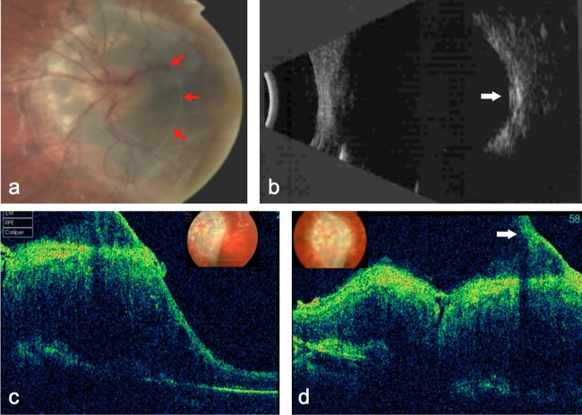
(A) Colour photograph of the posterior pole showed a grey elevated lesion involving the disc and macula with tortuous blood vessels. An overlying translucent fibrotic membrane was seen extending from 12–6 o'clock in a crescent configuration (red arrows). (B) B-scan ultrasonography showed the lesion to be slightly elevated (by 1 mm) with medium-to-high reflectivity (arrow). (C and D) Optical coherence tomography sections through the macula and disc, respectively, revealed thickened retina with loss of architectural layers, and a pre-retinal membrane causing vitreoretinal traction (arrow).
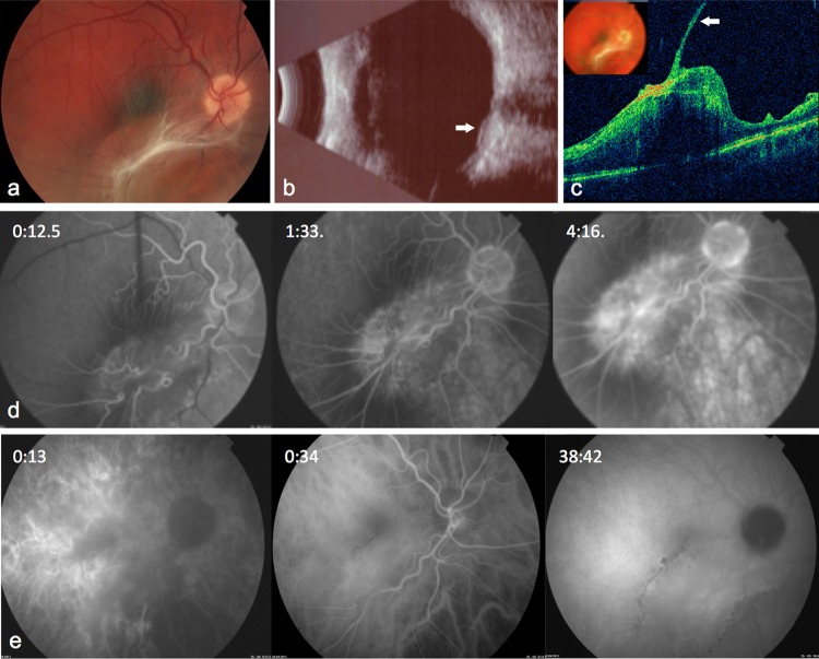
(A) Colour photograph of the right posterior pole showed an elevated lesion involving the inferior macula with a hyperpigmented margin which blends into the surrounding tissue. A grey-white fibrotic thickening is present over the surface of the lesion. (B) B-scan showed the lesion (arrow) to be slightly elevated with low-to-medium reflectivity. (C) Optical coherence tomography section through the centre of the lesion showed grossly thickened and disorganised retina with a tractional pre-retinal membrane (arrow). (D) Fundus fluorescein angiography demonstrated blockage of choroidal fluorescence by the outer pigmented portion of the lesion. Tortuosity of vessels within the lesion was prominent in the arterial phase. There was patchy dye leakage within the lesion and late staining. (E) Indocyanine green (ICG) did not reveal any significant choroidal vascular abnormality. Mild hyperfluorescence corresponding to the lesion and hypofluorescence outlining its border was seen in the late phase.
Similar articles
-
Retinal fluorescein and indocyanine green angiography and spectral-domain optical coherence tomography findings in acute retinal pigment epitheliitis.Retina. 2011 Jun;31(6):1156-63. doi: 10.1097/IAE.0b013e3181fbcea5. Retina. 2011. PMID: 21293312
-
[Diagnosis and differential diagnosis of the combined hamartoma of the retina and retinal pigment epithelium].Klin Monbl Augenheilkd. 2001 Nov;218(11):697-701. doi: 10.1055/s-2001-18660. Klin Monbl Augenheilkd. 2001. PMID: 11731896 German.
-
MultiColorTM imaging in combined hamartoma of the retina and retinal pigment epithelium.Eye (Lond). 2018 Sep;32(9):1478-1482. doi: 10.1038/s41433-018-0123-2. Epub 2018 May 23. Eye (Lond). 2018. PMID: 29789660 Free PMC article.
-
Combined hamartoma of sensory retina and retinal pigment epithelium.Retina. 1989;9(4):302-11. doi: 10.1097/00006982-198909040-00011. Retina. 1989. PMID: 2697919 Review.
-
Role of optical coherence tomography angiography in retinal tumors: A narrative review.Indian J Ophthalmol. 2024 Aug 1;72(8):1082-1090. doi: 10.4103/IJO.IJO_29_24. Epub 2024 Jul 29. Indian J Ophthalmol. 2024. PMID: 39078951 Free PMC article. Review.
Cited by
-
[Retinal tumors in adults: Part 2 nonvascular tumors of retina and retinal pigment epithelium].Ophthalmologe. 2021 Nov;118(11):1153-1160. doi: 10.1007/s00347-021-01446-w. Epub 2021 Jul 29. Ophthalmologe. 2021. PMID: 34327607 Review. German.
-
Combined hamartoma of the retina and retinal pigment epithelium - MRI features of a rare paediatric intraocular tumour.BJR Case Rep. 2020 Nov 17;7(2):20200077. doi: 10.1259/bjrcr.20200077. eCollection 2021 Apr 1. BJR Case Rep. 2020. PMID: 33841897 Free PMC article.
References
-
- Ting TD, McCuen BW, II, Fekrat S. Combined hamartoma of the retina and retinal pigment epithelium: optical coherence tomography. Retina 2002;22:98–101. - PubMed
-
- Font RL, Moura RA, Sheltar DJ, et al. Combined hamartoma of the sensory retina and retinal pigment epithelium. Retina 1989;9:302–11. - PubMed
-
- Shields CL, Thangappan A, Hartzell K, et al. Combined hamartoma of the retina and retinal pigment epithelium in 77 consecutive patients visual outcome based on macular versus extramacular tumor location. Ophthalmology 2008;115:2246–52. - PubMed
-
- Moschos M, Ladas ID, Zafirakis PK, et al. Recurrent vitreous hemorrhages due to combined pigment epithelial and retinal hamartoma: natural course and indocyanine green angiographic findings. Ophthalmologica 2001;215:66–9. - PubMed
Publication types
MeSH terms
Substances
LinkOut - more resources
Full Text Sources
Miscellaneous