

Impact of Partial and complete rupture of anterior cruciate ligament on medial meniscus: A cadavaric study
- PMID: 23162142
- PMCID: PMC3491783
- DOI: 10.4103/0019-5413.101040
Impact of Partial and complete rupture of anterior cruciate ligament on medial meniscus: A cadavaric study
Abstract
Background: The clinical relationship between medial meniscus tear and anterior cruciate ligament (ACL) rupture has been well documented. However, the mechanism of this clinical phenomenon is not exactly explained. Our aim is to investigate the biomechanical impact of partial and complete ACL rupture on different parts of medial meniscus.
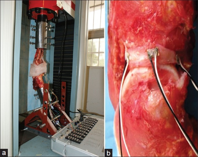
Materials and methods: TWELVE FRESH HUMAN CADAVERIC KNEE SPECIMENS WERE DIVIDED INTO FOUR GROUPS: ACL intact (ACL-I), anteromedial bundle transection (AMB-T), posterolateral bundle transection (PLB-T), and ACL complete transection (ACL-T) group. Strain on the anterior horn, body part, and posterior horn of medial meniscus were measured under 200 N axial compressive tibial load at 0°, 30°, 60°, and 90° of knee flexion, respectively.
Results: Compared with the control group (ACL-I), the ACL-T group had a higher strain on whole medial meniscus at 0°, 60°, and 90° of flexion. But at 30°, it had a higher strain on posterior horn of meniscus only. As to PLB-T group, strain on whole meniscus increased at full extension, while strain increased on posterior horn at 30° and on body of meniscus at 60°. However, AMB-T only brought about higher strain at 60° of flexion on body and posterior horn of meniscus.
Conclusions: Similar to complete rupture, partial rupture of ACL can also trigger strain concentration on medial meniscus, especially posterior horn, which may be a more critical reason for meniscus injury associated with chronic ACL deficiency.
Keywords: Anterior cruciate ligament; anterior cruciate ligament rupture; biomechanics; medial meniscal tear.
Conflict of interest statement
Figures


Similar articles
-
Effect of partial and complete posterior cruciate ligament transection on medial meniscus: A biomechanical evaluation in a cadaveric model.Indian J Orthop. 2013 Sep;47(5):493-9. doi: 10.4103/0019-5413.118206. Indian J Orthop. 2013. PMID: 24133310 Free PMC article.
-
Longitudinal tear of the medial meniscus posterior horn in the anterior cruciate ligament-deficient knee significantly influences anterior stability.Am J Sports Med. 2011 Oct;39(10):2187-93. doi: 10.1177/0363546511416597. Epub 2011 Aug 9. Am J Sports Med. 2011. PMID: 21828365
-
Partial meniscectomy does not affect the biomechanics of anterior cruciate ligament reconstructed knee with a lateral posterior meniscal root tear.Knee Surg Sports Traumatol Arthrosc. 2020 Nov;28(11):3481-3487. doi: 10.1007/s00167-020-06209-9. Epub 2020 Sep 5. Knee Surg Sports Traumatol Arthrosc. 2020. PMID: 32889558
-
[Biomechanical effect of anterior cruciate ligament rupture on posterior horn of lateral meniscus].Zhongguo Xiu Fu Chong Jian Wai Ke Za Zhi. 2010 Jan;24(1):14-6. Zhongguo Xiu Fu Chong Jian Wai Ke Za Zhi. 2010. PMID: 20135963 Chinese.
-
Posterior Medial Meniscus Root Tears Potentiate the Effect of Increased Tibial Slope on Anterior Cruciate Ligament Graft Forces.Am J Sports Med. 2020 Feb;48(2):334-340. doi: 10.1177/0363546519889628. Epub 2019 Dec 10. Am J Sports Med. 2020. PMID: 31821011
Cited by
-
Projecting Lifetime Risk of Symptomatic Knee Osteoarthritis and Total Knee Replacement in Individuals Sustaining a Complete Anterior Cruciate Ligament Tear in Early Adulthood.Arthritis Care Res (Hoboken). 2017 Feb;69(2):201-208. doi: 10.1002/acr.22940. Epub 2016 Dec 31. Arthritis Care Res (Hoboken). 2017. PMID: 27214559 Free PMC article.
-
Strong association of meniscus tears with complete Anterior Cruciate Ligament (ACL) injuries relative to partial ACL injuries.J Clin Orthop Trauma. 2021 Oct 26;23:101671. doi: 10.1016/j.jcot.2021.101671. eCollection 2021 Dec. J Clin Orthop Trauma. 2021. PMID: 34790561 Free PMC article.
-
Effect of partial and complete posterior cruciate ligament transection on medial meniscus: A biomechanical evaluation in a cadaveric model.Indian J Orthop. 2013 Sep;47(5):493-9. doi: 10.4103/0019-5413.118206. Indian J Orthop. 2013. PMID: 24133310 Free PMC article.
-
Subchondral injection of human umbilical cord mesenchymal stem cells ameliorates knee osteoarthritis by inhibiting osteoblast apoptosis and TGF-beta activity.Stem Cell Res Ther. 2025 May 9;16(1):235. doi: 10.1186/s13287-025-04366-7. Stem Cell Res Ther. 2025. PMID: 40346614 Free PMC article.
-
Delay in surgery predisposes to meniscal and chondral injuries in anterior cruciate ligament deficient knees.Indian J Orthop. 2016 Sep;50(5):492-498. doi: 10.4103/0019-5413.189606. Indian J Orthop. 2016. PMID: 27746491 Free PMC article.
References
-
- Henning CE. Current status of meniscus salvage. Clin Sports Med. 1990;9:567–76. - PubMed
-
- Yoon KH, Yoo JH, Kim KI. Bone contusion and associated meniscal and medial collateral ligament injury in patients with anterior cruciate ligament rupture. J Bone Joint Surg Am. 2011;93:1510–8. - PubMed
-
- Naranje S, Mittal R, Nag H, Sharma R. Arthroscopic and magnetic resonance imaging evaluation of meniscus lesions in the chronic anterior cruciate ligament-deficient knee. Arthroscopy. 2008;24:1045–51. - PubMed
-
- Panisset JC, Duraffour H, Vasconcelos W, Colombet P, Javois C, Potel JF, et al. Société française d’arthroscopie. Clinical, radiological and arthroscopic analysis of the ACL tear. A prospective study of 418 cases. Rev Chir Orthop Reparatrice Appar Mot. 2008;94(8 Suppl):362–8. - PubMed
-
- Allen CR, Wong EK, Livesay GA, Sakane M, Fu FH, Woo SL. Importance of the medial meniscus in the anterior cruciate ligament-deficient knee. J Orthop Res. 2000;18:109–15. - PubMed
Grants and funding
LinkOut - more resources
Full Text Sources
Research Materials