

The management of perineal wounds
- PMID: 23162235
- PMCID: PMC3495386
- DOI: 10.4103/0970-0358.101318
The management of perineal wounds
Abstract
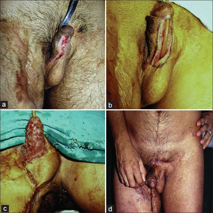
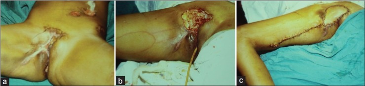
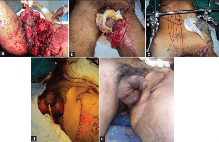
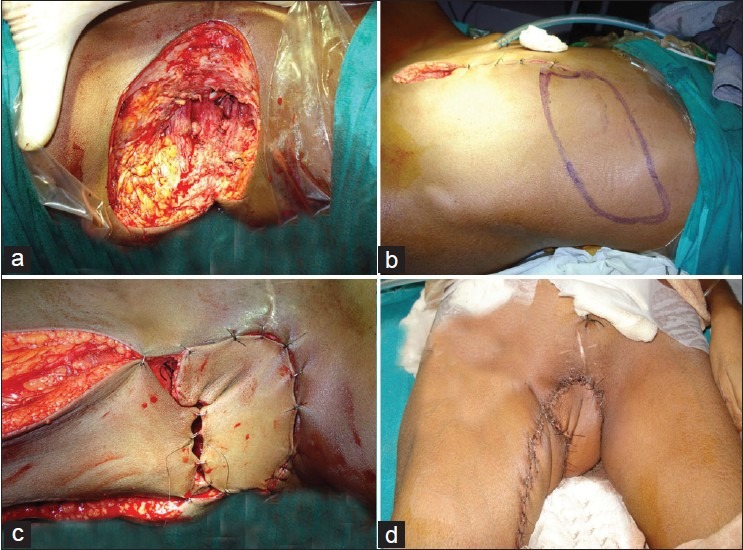
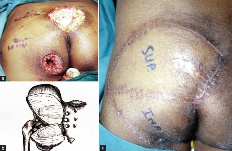
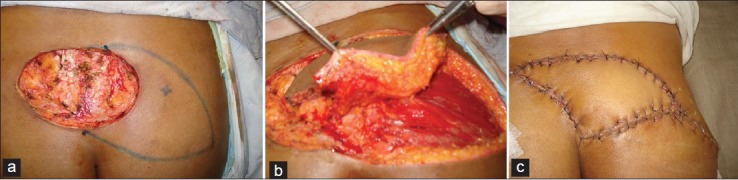
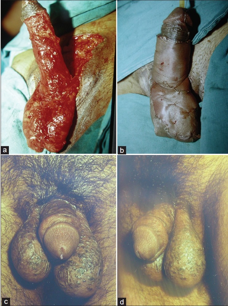
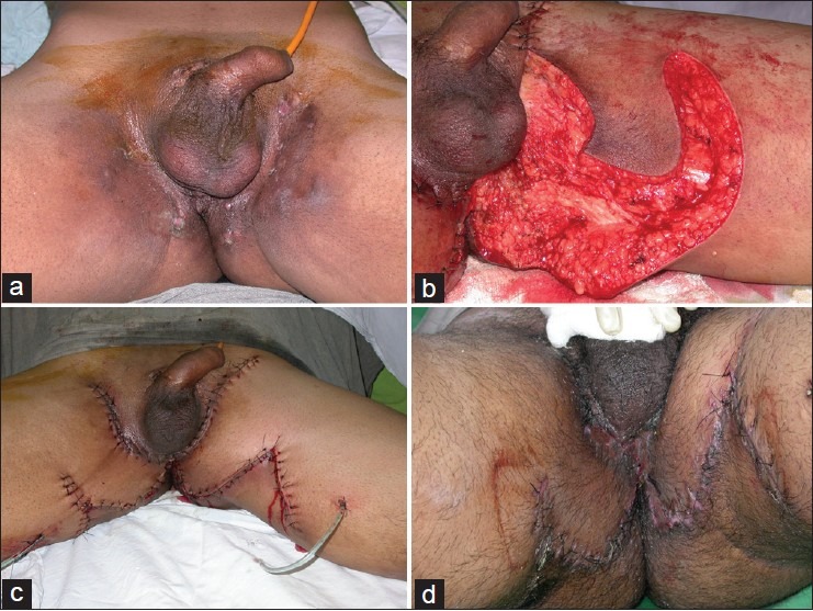
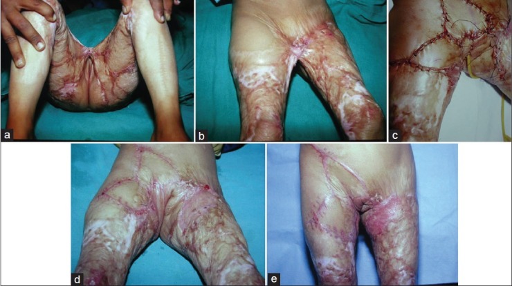
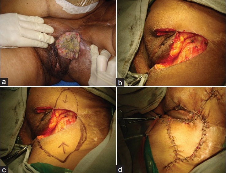
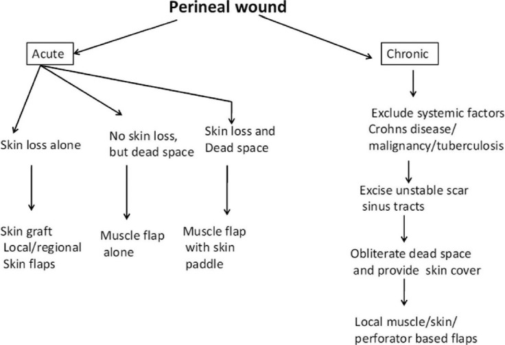
Management of perineal wounds can be very frustrating as these invariably get contaminated from the ano-genital tracts. Moreover, the apparent skin defect may be associated with a significant three dimensional dead space in the pelvic region. Such wounds are likely to become chronic and recalcitrant if appropriate wound management is not instituted in a timely manner. These wounds usually result after tumor excision, following trauma or as a result of infective pathologies like hideradenitis suppurativa or following thermal burns. Many options are available for management of perineal wounds and these have been discussed with illustrative case examples. A review of literature has been done for listing commonly instituted options for management of the wounds in perineum.
Keywords: Muscle flaps; perforator flaps; perineum; reconstructive options; skin grafts; vacuum assisted.
Conflict of interest statement
Figures
Similar articles
-
The Free-style Gluteal Perforator Flap in the Thinning and Delay Process for Perineal Reconstruction After Necrotizing Fasciitis.Wounds. 2016 Jun;28(6):200-5. Wounds. 2016. PMID: 27434419
-
Superior and inferior gluteal artery perforator flaps in reconstruction of gluteal and perianal/perineal hidradenitis suppurativa lesions.Microsurgery. 2011 Oct;31(7):539-44. doi: 10.1002/micr.20918. Epub 2011 Aug 23. Microsurgery. 2011. PMID: 21976179
-
The management of burns to the perineum and genitals.J Burn Care Rehabil. 1990 Jan-Feb;11(1):54-6. doi: 10.1097/00004630-199001000-00012. J Burn Care Rehabil. 1990. PMID: 2312592
-
Reconstruction of perineal defects.Ann R Coll Surg Engl. 2013 Nov;95(8):539-44. doi: 10.1308/rcsann.2013.95.8.539. Ann R Coll Surg Engl. 2013. PMID: 24165333 Free PMC article. Review.
-
[Perineal injuries in wartime].Ann Urol (Paris). 1997;31(5):303-8. Ann Urol (Paris). 1997. PMID: 9480637 Review. French.
Cited by
-
Wound Infection After Keloid Excision and Adjuvant Radiotherapy: Two Case Reports and Literature Review.Clin Cosmet Investig Dermatol. 2025 Aug 13;18:1943-1951. doi: 10.2147/CCID.S545846. eCollection 2025. Clin Cosmet Investig Dermatol. 2025. PMID: 40827224 Free PMC article.
-
How to Treat a Cyclist's Nodule?-Introduction of a Novel, ICG-Assisted Approach.J Clin Med. 2024 Feb 16;13(4):1124. doi: 10.3390/jcm13041124. J Clin Med. 2024. PMID: 38398438 Free PMC article.
-
Gracilis Myocutaneous Flap: Adding to the Armamentarium of Complex Sacrococcygeal Defect Reconstruction.Indian J Plast Surg. 2019 May;52(2):246-249. doi: 10.1055/s-0039-1696078. Epub 2019 Sep 27. Indian J Plast Surg. 2019. PMID: 31602144 Free PMC article.
-
Outcomes of gracilis muscle interposition for rectourethral fistulas caused by treatment of prostate cancer.Tech Coloproctol. 2023 Oct;27(10):937-944. doi: 10.1007/s10151-023-02759-5. Epub 2023 Feb 17. Tech Coloproctol. 2023. PMID: 36800073
-
Perineal bull gore with urinary bladder perforation and pneumoperitoneum.J Clin Diagn Res. 2013 May;7(5):902-4. doi: 10.7860/JCDR/2013/5556.2981. Epub 2013 Mar 22. J Clin Diagn Res. 2013. PMID: 23814738 Free PMC article.
References
-
- [visited on 18 04 2012 at 7pm]. http://www.merriam-webster.com/dictionary/perineum .
-
- McAllister E, Wells K, Chaet M, Norman J, Cruse W. Perineal reconstruction after surgical extirpation of pelvic malignancies using the transpelvic transverse rectus abdominus musculocutaneous flap. Ann Surg Oncol. 1994;1:164–8. - PubMed
-
- Yeh KA, Hoffman JP, Kusiak JE, Litwin S, Sigurdson ER, Eisenberg BL. Reconstruction with musculocutaneous flaps following resection of locally recurrent rectal cancer. Am Surg. 1995;61:581–9. - PubMed
-
- Touran T, Frost DB, O’Connell TX. Sacral resection: Operative technique and outcome. Arch Surg. 1990;125:911–3. - PubMed
-
- McCraw JB, Massey FM, Shanklin KD, Horton CE. Vaginal reconstruction with gracilis musculocutaneous flaps. Plast Reconstr Surg. 1976;58:176–83. - PubMed
LinkOut - more resources
Full Text Sources