

doi: 10.1016/j.jsmc.2012.06.010.
Epub 2012 Sep 4.
Neuropharmacology of Sleep and Wakefulness: 2012 Update
Affiliations
- PMID: 23162386
- PMCID: PMC3496285
- DOI: 10.1016/j.jsmc.2012.06.010
Item in Clipboard
Neuropharmacology of Sleep and Wakefulness: 2012 Update
Sleep Med Clin.
.
Abstract
The development of sedative/hypnotic molecules has been empiric rather than rational. The empiric approach has produced clinically useful drugs but for no drug is the mechanism of action completely understood. All available sedative/hypnotic medications have unwanted side effects and none of these medications creates a sleep architecture that is identical to the architecture of naturally occurring sleep. This chapter reviews recent advances in research aiming to elucidate the neurochemical mechanisms regulating sleep and wakefulness. One promise of rational drug design is that understanding the mechanisms of sedative/hypnotic action will significantly enhance drug safety and efficacy.
Figures
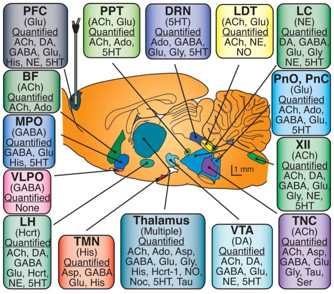
Sagittal drawing of the rat brain (modified from) schematizes the location, shape, and size of some brain regions that regulate sleep and wakefulness. The name of each brain region appears in bold print, the major neurotransmitters used for signaling to other brain regions are in parentheses, and neurochemical analytes relevant for arousal-state control that have been measured in that brain region are listed under the header “Quantified ”. The microdialysis probe is drawn to scale and is shown sampling from the prefrontal cortex. Abbreviations: XII – hypoglossal nucleus; BF – basal forebrain; DRN – dorsal raphé nucleus; LC – locus coeruleus; LDT – laterodorsal tegmental nucleus; LH – lateral hypothalamus; MPO – medial preoptic area; PFC – prefrontal cortex; PPT – pedunculopontine tegmental nucleus; PnC – pontine reticular formation, caudal part; PnO – pontine reticular formation, oral part; TMN – tuberomamillary nucleus; TNC – trigeminal nucleus complex; VLPO – ventrolateral preoptic area; VTA – ventral tegmental area; 5HT – serotonin; ACh – acetylcholine; Ado – adenosine; Asp – aspartate; DA – dopamine; GABA – γ-aminobutyric acid; Glu – glutamate; Gly – glycine; His – histamine; Hcrt – hypocretin; NE – norepinephrine; NO – nitric oxide; Noc – nociceptin; Ser – serine; 5HT – serotonin; Tau – taurine. Figure reprinted from Watson et al., 2010 with permission.
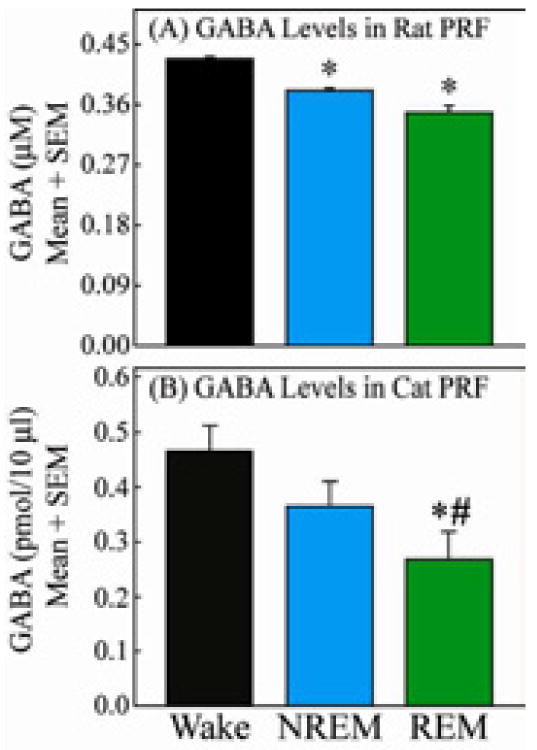
These comparative data illustrate two key points. First, that in both rat (A) and cat (B) there are parallel, state-dependent changes in GABA levels. In rat and cat GABA levels are significantly lower in REM sleep than during wakefulness. Second, methodological differences in the collection of GABA preclude direct comparison of GABA levels between these two species. GABA levels shown in A and B reflect differences in microdialysis flowrate (0.4 μL/min for rat and 2.0 μL/min for cat), molecular weight cut-off of the microdialysis probe membrane (18000 Daltons for rat and 6 Daltons for cat) and possibly membrane material (regenerated cellulose for rat and cuprophane for cat). Figures modified from Watson et al., 2011 and Vanini et al., 2011 with permission.
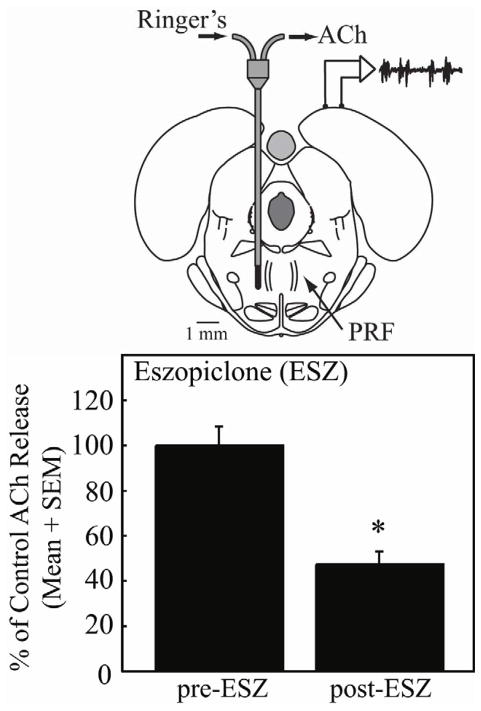
Top: schematic coronal section of rat brain stem illustrates placement of a microdialysis probe in the PRF. Ringer’s solution is pumped into the probe and samples are collected for quantification of ACh. Schematized at top right of brain are electrodes and an amplifier for recording the cortical electroencephalogram (EEG), and a representative trace showing EEG activity after intravenous administration of eszopiclone. Bottom: Histograms summarize the significant decrease in ACh release within the PRF caused by intravenous administration of eszopiclone. Data reprinted from Hambrecht-Wiedbusch et al., 2010 with permission.
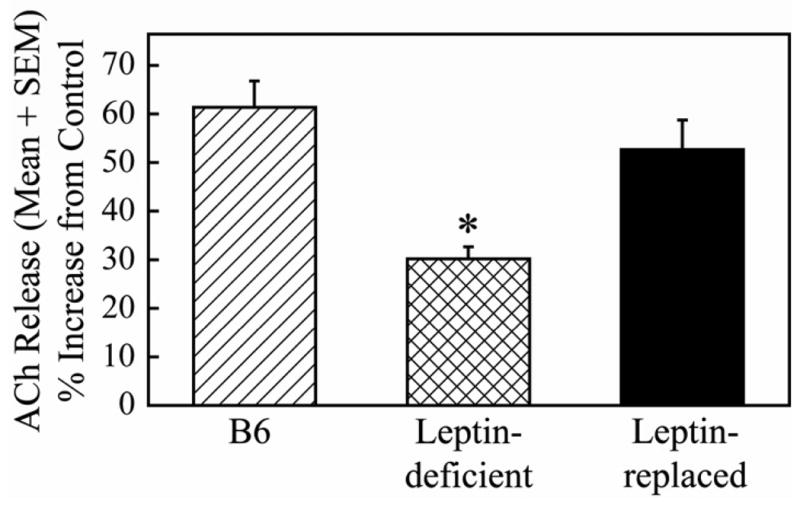
Dialysis administration of olanzapine (100 μM) to the prefrontal cortex of C57BL/6J (B6), leptin-deficient, or leptin-replaced mice caused an increase in ACh release in the prefrontal cortex. The increase in ACh release was significantly smaller in leptin-deficient mice compared to B6 controls. The olanzanpine-induced increase in ACh release was not significantly different between B6 controls and leptin-replaced mice. This suggests that leptin modulates the release of ACh within the prefrontal cortex and may also play a role in the cortical activation that occurs during wakefulness and REM sleep. Data reprinted from Wathen et al., 2012 with permission.
Similar articles
-
Neuropharmacology of Sleep and Wakefulness.Sleep Med Clin. 2010 Dec;5(4):513-528. doi: 10.1016/j.jsmc.2010.08.003. Sleep Med Clin. 2010. PMID: 21278831 Free PMC article.
-
An Update on the Use of Sedative-Hypnotic Medications in Psychiatric Disorders.Curr Psychiatry Rep. 2016 Sep;18(9):78. doi: 10.1007/s11920-016-0717-y. Curr Psychiatry Rep. 2016. PMID: 27417512 Free PMC article. Review.
-
Pinelliae Rhizoma Praeparatum Cum Alumine Extract: Sedative and Hypnotic Effects in Mice and Component Compounds.Biomed Res Int. 2019 Apr 28;2019:6198067. doi: 10.1155/2019/6198067. eCollection 2019. Biomed Res Int. 2019. PMID: 31183370 Free PMC article.
-
Effects of sleep disorders and sedative-hypnotic medications on health-related quality of life in dialysis patients.Int Urol Nephrol. 2019 Jan;51(1):163-174. doi: 10.1007/s11255-018-2018-3. Epub 2018 Nov 22. Int Urol Nephrol. 2019. PMID: 30467784
-
Pharmacological advances in the treatment of insomnia.Curr Pharm Des. 2011;17(15):1471-5. doi: 10.2174/138161211796197052. Curr Pharm Des. 2011. PMID: 21476952 Review.
Cited by
-
Effects of interface pressure distribution on human sleep quality.PLoS One. 2014 Jun 12;9(6):e99969. doi: 10.1371/journal.pone.0099969. eCollection 2014. PLoS One. 2014. PMID: 24924427 Free PMC article. Clinical Trial.
-
Depression of Non-Neuronal Cholinergic System May Play a Role in Co-Occurrence of Subjective Daytime Sleepiness and Hypertension in Patients with Obstructive Sleep Apnea Syndrome.Nat Sci Sleep. 2021 Dec 14;13:2153-2163. doi: 10.2147/NSS.S339038. eCollection 2021. Nat Sci Sleep. 2021. PMID: 34934375 Free PMC article.
-
Partial normalization of hippocampal oscillatory activity during sleep in TgF344-AD rats coincides with increased cholinergic synapses at early-plaque stage of Alzheimer's disease.Acta Neuropathol Commun. 2025 May 10;13(1):96. doi: 10.1186/s40478-025-02016-w. Acta Neuropathol Commun. 2025. PMID: 40349073 Free PMC article.
-
Ghrelin mediated regulation of neurosynaptic transmitters in depressive disorders.Curr Res Pharmacol Drug Discov. 2022 Jun 13;3:100113. doi: 10.1016/j.crphar.2022.100113. eCollection 2022. Curr Res Pharmacol Drug Discov. 2022. PMID: 35782191 Free PMC article. Review.
-
Variations in loop gain and arousal threshold during NREM sleep are affected by time of day over a 24-hour period in participants with obstructive sleep apnea.J Appl Physiol (1985). 2020 Oct 1;129(4):800-809. doi: 10.1152/japplphysiol.00376.2020. Epub 2020 Aug 13. J Appl Physiol (1985). 2020. PMID: 32790595 Free PMC article.
References
-
- Watson CJ, Baghdoyan HA, Lydic R. A neurochemical perspective on states of consciousness. In: Hudetz AG, Pearce RA, editors. Suppressing the Mind: Anesthetic Modulation of Memory and Consciousness. New York: Springer/Humana Press; 2010. pp. 33–80.
-
- Steriade M, McCarley RW, editors. Brain Control of Wakefulness and Sleep. New York: Kluwer Academic/Plenum Publishers; 2005.
-
- McCarley RW. Neurobiology of REM and NREM sleep. Sleep Med. 2007;8(4):302–330. - PubMed
Grants and funding
LinkOut - more resources
Full Text Sources
