. 2012 Nov 14:6:165.
doi: 10.3389/fnins.2012.00165. eCollection 2012.

Inhibitory control of hippocampal inhibitory neurons

Affiliations

Inhibitory control of hippocampal inhibitory neurons

Simon Chamberland et al. Front Neurosci. .

Abstract

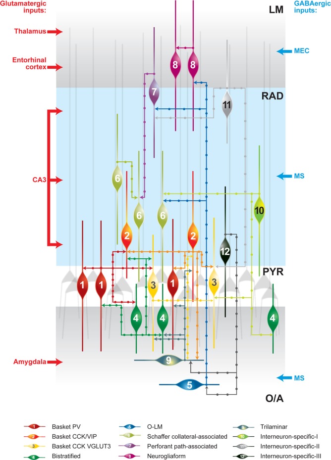
Information processing within neuronal networks is determined by a dynamic partnership between principal neurons and local circuit inhibitory interneurons. The population of GABAergic interneurons is extremely heterogeneous and comprises, in many brain regions, cells with divergent morphological and physiological properties, distinct molecular expression profiles, and highly specialized functions. GABAergic interneurons have been studied extensively during the past two decades, especially in the hippocampus, which is a relatively simple cortical structure. Different types of hippocampal inhibitory interneurons control spike initiation [e.g., axo-axonic and basket cells (BCs)] and synaptic integration (e.g., bistratified and oriens-lacunosum moleculare interneurons) within pyramidal neurons and synchronize local network activity, providing a means for functional segregation of neuronal ensembles and proper routing of hippocampal information. Thus, it is thought that, at least in the hippocampus, GABAergic inhibitory interneurons represent critical regulating elements at all stages of information processing, from synaptic integration and spike generation to large-scale network activity. However, this raises an important question: if inhibitory interneurons are fundamental for network computations, what are the mechanisms that control the activity of the interneurons themselves? Given the essential role of synaptic inhibition in the regulation of neuronal activity, it would be logical to expect that specific inhibitory mechanisms have evolved to control the operation of interneurons. Here, we review the mechanisms of synaptic inhibition of interneurons and discuss their role in the operation of hippocampal inhibitory circuits.

Keywords: GABA; hippocampus; inhibition; interneuron-specific interneuron; synapse.


Figures

Figure 1
Figure 1
Schematic representation of synaptically connected GABAergic inhibitory circuits in the CA1 hippocampal area. The main glutamatergic inputs are indicated on the left. The main long-range GABAergic projections contacting CA1 interneurons are shown on the right. Some connections are indicated on the basis of data from one recording and further analysis may be required. CCK, cholecystokinin; LM, stratum lacunosum moleculare; O/A, stratum oriens/alveus; O–LM, oriens–lacunosum moleculare cell; PV, parvalbumin; PYR, stratum pyramidale; RAD, stratum radiatum; VGLUT3, vesicular glutamate transporter 3; VIP, vasoactive intestinal peptide.
Figure 2
Figure 2
VIP-positive interneurons at the PYR/RAD border target O–LM interneurons. (A) Maximal projection of a two-photon z-stack acquired in the CA1 region of the hippocampus of a VIP-eGFP mouse, showing bipolarly oriented VIP-positive cell bodies located at the PYR/RAD border and a dense axonal arborization in the O/A. (B) Immunofluorescence images of neurons located in PYR positive for calretinin (top) and VIP (middle) as well as their superimposition (bottom). Scale bar: 20 μ m. (C) Reconstruction of a bipolarly oriented VIP-positive cell, showing anatomical features of IS-IIIs (soma and dendrites are shown in black and axon is shown in red) and its irregularly spiking firing pattern typical for these cells. (D) Neurolucida reconstruction of a connected pair of interneurons: presynaptic IS-III (soma and dendrites are in black and axon is in red) and postsynaptic O–LM (soma and dendrites are in green, axon is in blue) and examples of unitary IPSCs evoked by two-photon glutamate uncaging (bottom left) and presynaptic spikes during paired recordings (bottom right). Black arrows indicate three putative contact sites onto O–LM dendrites. Modified from Chamberland et al. (2010).

References

    1. Acsady L., Arabadzisz D., Freund T. F. (1996). Correlated morphological and neurochemical features identify different subsets of vasoactive intestinal polypeptide-immunoreactive interneurons in rat hippocampus. Neuroscience 73, 299–315 10.1016/0306-4522(95)00610-9 - DOI - PubMed
    1. Acsady L., Gorcs T. J., Freund T. F. (1996). Different populations of vasoactive intestinal polypeptide-immunoreactive interneurons are specialized to control pyramidal cells or interneurons in the hippocampus. Neuroscience 73, 317–334 10.1016/0306-4522(95)00609-5 - DOI - PubMed
    1. Ali A. B. (2007). Presynaptic Inhibition of GABAA receptor-mediated unitary IPSPs by cannabinoid receptors at synapses between CCK-positive interneurons in rat hippocampus. J. Neurophysiol. 98, 861–869 10.1152/jn.00156.2007 - DOI - PubMed
    1. Ali A. B. (2011). CB1 modulation of temporally distinct synaptic facilitation among local circuit interneurons mediated by N-type calcium channels in CA1. J. Neurophysiol. 105, 1051–1062 10.1152/jn.00831.2010 - DOI - PMC - PubMed
    1. Ali A. B., Bannister A. P., Thomson A. M. (1999). IPSPs elicited in CA1 pyramidal cells by putative basket cells in slices of adult rat hippocampus. Eur. J. Neurosci. 11, 1741–1753 10.1046/j.1460-9568.1999.00592.x - DOI - PubMed