. 2012 Nov 16:6:84.
doi: 10.3389/fncir.2012.00084. eCollection 2012.

Connectivity from OR37 expressing olfactory sensory neurons to distinct cell types in the hypothalamus

Affiliations

Connectivity from OR37 expressing olfactory sensory neurons to distinct cell types in the hypothalamus

Andrea Bader et al. Front Neural Circuits. .

Abstract

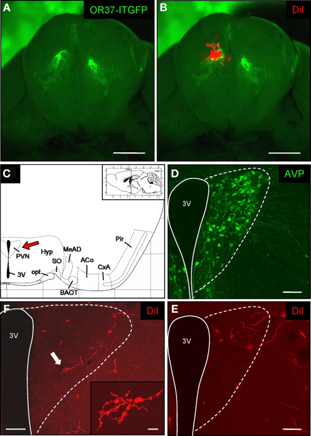
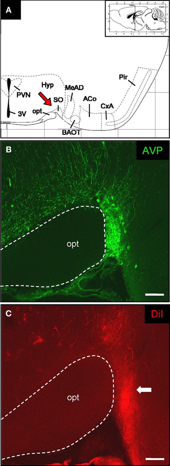
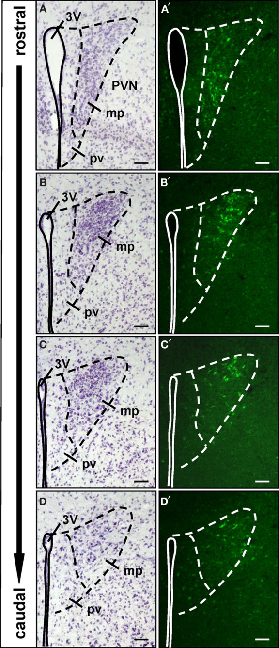
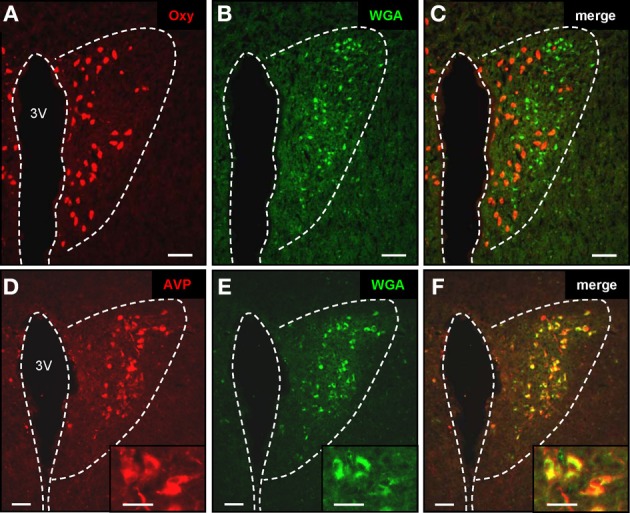
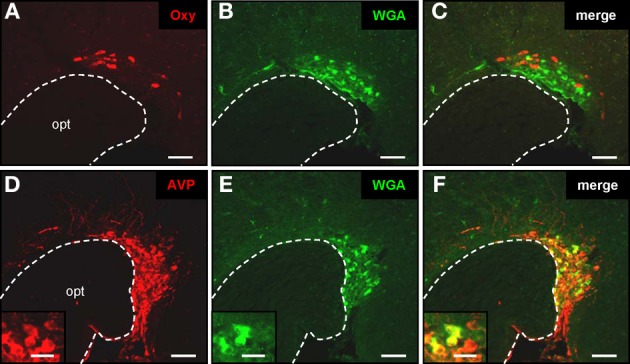
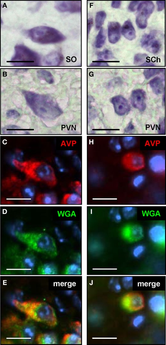
Olfactory sensory neurons (OSNs) which express a member from the OR37 subfamily of odorant receptor (OR) genes are wired to the main olfactory bulb (MOB) in a unique monoglomerular fashion; from these glomeruli an untypical connectivity into higher brain centers exists. In the present study we have investigated by DiI and transsynaptic tracing approaches how the connection pattern from these glomeruli into distinct hypothalamic nuclei is organized. The application of DiI onto the ventral domain of the bulb which harbors the OR37 glomeruli resulted in the labeling of fibers within the paraventricular nucleus (PVN) and supraoptic nucleus (SO) of the hypothalamus; some of these fibers were covered with varicose-like structures. No DiI-labeled cell somata were detectable in these nuclei. The data indicate that projection neurons which originate in the OR37 region of the MOB form direct connections into these nuclei. The cells that were labeled by the transsynaptic tracer WGA in these nuclei were further characterized. Their distribution pattern in the paraventricular nucleus was reminiscent of cells which produce distinct neuropeptides. Double labeling experiments confirmed that they contained vasopressin, but not the related neuropeptide oxytocin. Morphological analysis revealed that they comprise of magno- and parvocellular cells. A comparative investigation of the WGA-positive cells in the SO demonstrated that these were vasopressin-positive, as well, whereas oxytocin-producing cells of this nucleus also contained no transsynaptic tracer. Together, the data demonstrates a connectivity from OR37 expressing sensory neurons to distinct hypothalamic neurons with the same neuropeptide content.

Keywords: OR37; olfaction; paraventricular nucleus; supraoptic nucleus; vasopressin; wiring.


Figures

Figure 1
Figure 1
DiI-labeling in the paraventricular nucleus of the hypothalamus after tracer application onto the OR37 glomerulus. (A) Whole-mount preparation of the olfactory bulbs from an OR37-ITGFP mouse. A single fluorescent glomerulus (green) is visible on the ventral surface of each bulb. Scale bar: 1 mm. (B) A DiI crystal (red) was deposited on the fluorescent glomerulus. Scale bar: 1 mm. (C) Schematic representation of a section through the mouse brain [adapted from Paxinos and Franklin (2001)]. The position of the section along the anterior–posterior axis is indicated on the inset. The interval of the lattice is 1 mm. Several distinct nuclei are indicated. The red arrow highlights the paraventricular nucleus. 3V, third ventricle; ACo, anterior cortical amygdala; BAOT, bed nucleus of the accessory olfactory tract; CxA, cortex-amygdala transition zone; MeAD, medial amygdala anterior-dorsal part; opt, optic tract; Pir, piriform cortex; PVN, paraventricular nucleus; SO, supraoptic nucleus. (D) Confocal image of a tissue section through the paraventricular nucleus. Vasopressinergic cells are stained with a specific antibody (anti-neurophysin-vasopressin). According to this cell population the extent of the nucleus close to the third ventricle (3V) can be estimated (dotted lines) and is transferred to the neighboring section shown in (C). Pinhole size: 70 μm. Scale bar: 100 μm. (E) On the neighboring section distinct DiI-labeled fibers are located in the paraventricular nucleus after application of a DiI-crystal onto the OR37 glomerulus and subsequent incubation for 12 weeks. Pinhole size: 80 μm. Scale bar: 100 μm. (F) DiI-labeled fibers in the paraventricular nucleus of an individual 26 weeks after dye application. The arrow highlights the fiber shown on the inset. At high magnification, multiple ramifications and varicose-like swellings along the fiber are visible. Pinhole size: 80 μm; inset: 87 μm. Scale bar: 100 μm; inset: 10 μm.
Figure 2
Figure 2
DiI-labeled fibers in the supraoptic nucleus of the hypothalamus. (A) Schematic representation of a section through the mouse brain [adapted from Paxinos and Franklin (2001)]. The position of the section along the anterior–posterior axis is indicated on the inset. The interval of the lattice is 1 mm. Several distinct nuclei are indicated. The red arrow highlights the supraoptic nucleus. 3V, third ventricle; ACo, anterior cortical amygdala; BAOT, bed nucleus of the accessory olfactory tract; CxA, cortex-amygdala transition zone; MeAD: medial amygdala anterior-dorsal part; opt, optic tract; Pir, piriform cortex; PVN, paraventricular nucleus; SO: supraoptic nucleus. (B) Confocal image of a cross section through the supraoptic nucleus. Vasopressinergic cells are stained with a specific antibody (anti-neurophysin-vasopressin). According to this cell population the position of this particular hypothalamic nucleus close to the optic tract can be exactly determined. Pinhole size: 70 μm. Scale bar: 100 μm. (C) After application of DiI onto the OR37 glomerulus a prominent bundle of labeled fibers can be visualized on the neighboring tissue section. Comparing the two staining patterns shows that the DiI-labeled fibers extend up to the somatic portion of the supraoptic nucleus (arrow). Pinhole size: 80 μm. Scale bar: 100 μm.
Figure 3
Figure 3
Spatial organization of the WGA-immunoreactive cells in the paraventricular nucleus. (A–D) Nissl-stained cross sections through the paraventricular nucleus (PVN). The third ventricle (3V) is indicated by the continuous line. The dashed lines mark the extent of the paraventricular nucleus and separate the periventricular part (pv) from the major portion (mp) of the nucleus. These divisions are transferred to the respective neighboring sections shown in (A′–D′). (A′–D′) All along the anterior–posterior axis of the paraventricular nucleus, no WGA-immunoreactivity can be observed in the periventricular part. WGA-immunoreactive cells are located in the major portion of the nucleus. In the rostral part these cells are clustered in the ventromedial subdivision, at more caudal levels the WGA-immunoreactivity is predominantly located in dorsolateral parts. All pictures represent wide field images. The spacing between the sections from different areas along the anterior-posterior axis is approximately 60–96 μm, respectively. Scale bars: 50 μm.
Figure 4
Figure 4
Transsynaptic labeling of vasopressinergic neurons in the paraventricular nucleus. (A–C) Cross section through the paraventricular nucleus. By means of a specific anti-oxytocin antibody, multiple oxytonergic neurons can be visualized in close vicinity to the third ventricle (3V) (A; red). On the same section WGA-immunoreactive cells can be observed (B; green). Merging the two pictures (C) reveals that oxytonergic neurons are not labeled by the transsynaptic marker. Scale bars: 50 μm. (D–F) Tissue section through the paraventricular nucleus. After staining with an anti-vasopressin antibody, neurons are visible near the third ventricle (3V) (A; red). WGA-immunoreactive cells are located on the same section (B; green). Merge of pictures (A) and (B) reveals that vasopressinergic cells are labeled by the transsynaptic marker (C; yellow). Scale bars: 50 μm. Higher magnifications are shown on the insets. Scale bars: 25 μm. All pictures represent wide field images.
Figure 5
Figure 5
Transsynaptic labeling of vasopressinergic neurons in the supraoptic nucleus. (A–C) Cross section through the supraoptic nucleus. By means of a specific anti-oxytocin antibody, multiple oxytonergic neurons can be visualized near the optic tract (opt) (A; red). On the same section WGA-immunoreactive cells can be observed (B; green). Merging the two pictures (C) reveals that oxytonergic neurons are not labeled by the transsynaptic marker. Scale bars: 50 μm. (D–F) Tissue section through the supraoptic nucleus. After immunohistochemical staining with an anti-neurophysin-vasopressin antibody, neurons are visible in close vicinity to the optic tract (opt) (A; red). On the same section WGA-immunoreactive cells are located (B; green). Merge of pictures (A) and (B) reveals that vasopressinergic cells are labeled by the transsynaptic marker (C; yellow). Scale bars: 50 μm. Higher magnifications are shown on the insets. Scale bars: 20 μm. All pictures represent wide field images.
Figure 6
Figure 6
Magno- and parvocellular vasopressinergic neurons in the paraventricular nucleus are labeled by the transsynaptic marker. (A) Nissl staining of a section through the supraoptic nucleus (SO) reveals the typical morphology of magnocellular neurons; these cells are large in diameter and exhibit a spindle-shaped morphology. (B) On Nissl stained sections through the paraventricular nucleus (PVN), cells with a similar morphology as shown in (A), thus magnocellular neurons, can be identified. (C) Section through the paraventricular nucleus after incubation with an anti-AVP antibody; neurons with a typical magnocellular morphology are stained (D) Section shown in (C) after incubation with an anti-WGA antibody. (E) The overlay of pictures (C) and (D) shows that this magnocellular vasopressinergic neuron is also labeled by the transsynaptic marker. (F) Nissl staining of a section through the suprachiasmatic nucleus (SCh) reveals the typical morphology of parvocellular neurons; these cells have a small diameter and are round-shaped. (G) On Nissl stained sections through the paraventricular nucleus (PVN), cells with a similar morphology as shown in (F), thus parvocellular neurons, can be identified. (H) Section through the paraventricular nucleus after incubation with an anti-AVP antibody; neurons with a typical parvocellular morphology are stained. (I) Section shown in (H) after incubation with an anti-WGA antibody. (J) The overlay of pictures (H) and (I) shows that this parvocellular vasopressinergic neuron is also labeled by the transsynaptic marker. All pictures represent wide field images. Scale bars: 10 μm

Similar articles

Cited by

References

    1. Albers H. E. (2012). The regulation of social recognition, social communication and aggression: vasopressin in the social behavior neural network. Horm. Behav. 61, 283–292 10.1016/j.yhbeh.2011.10.007 - DOI - PubMed
    1. Almeida O., Gozdowska M., Kulczykowska E., Oliveira R. F. (2012). Brain levels of arginine-vasotocin and isotocin in dominant and subordinate males of a cichlid fish. Horm. Behav. 61, 212–217 10.1016/j.yhbeh.2011.12.008 - DOI - PubMed
    1. Antoni F. A. (1993). Vasopressinergic control of pituitary adrenocorticotropin secretion comes of age. Front. Neuroendocrinol. 14:4 10.1006/frne.1993.1004 - DOI - PubMed
    1. Bader A., Breer H., Strotmann J. (2012). Untypical connectivity from olfactory sensory neurons expressing OR37 into higher brain centers visualized by genetic tracing. Histochem. Cell Biol. 137, 615–628 10.1007/s00418-012-0919-2 - DOI - PubMed
    1. Bautze V., Bär R., Fissler B., Trapp M., Schmidt D., Beifuss U., et al. (2012). Mammalian-specific OR37 receptors are differentially activated by distinct odorous fatty aldehydes. Chem. Senses 37, 479–493 10.1093/chemse/bjr130 - DOI - PubMed

LinkOut - more resources