

Abstract art and cortical motor activation: an EEG study
- PMID: 23162456
- PMCID: PMC3499799
- DOI: 10.3389/fnhum.2012.00311
Abstract art and cortical motor activation: an EEG study
Abstract
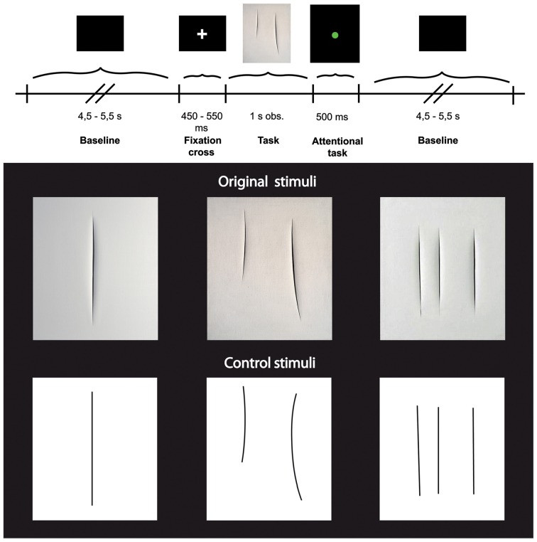
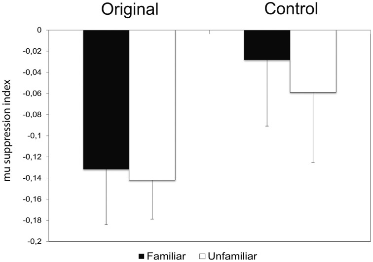
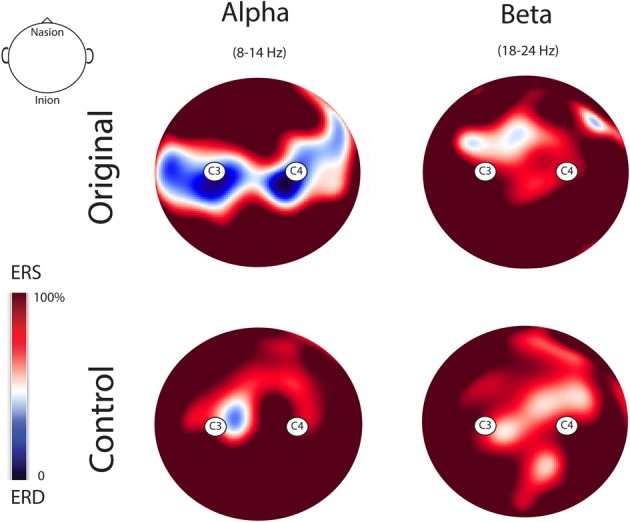
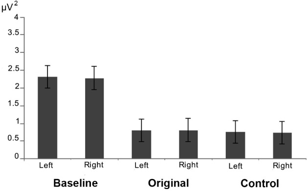
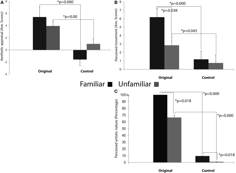
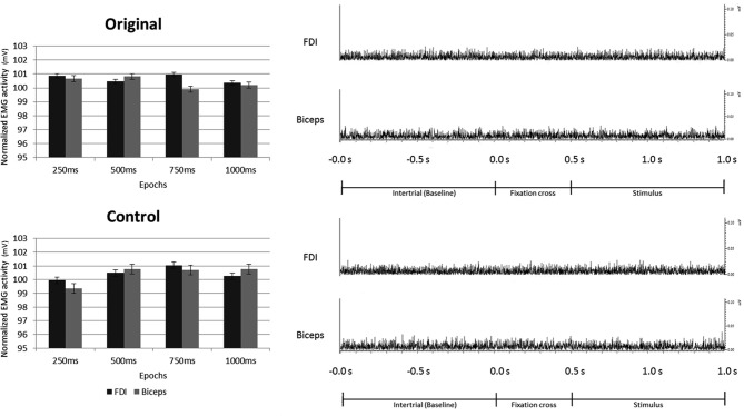
The role of the motor system in the perception of visual art remains to be better understood. Earlier studies on the visual perception of abstract art (from Gestalt theory, as in Arnheim, 1954 and 1988, to balance preference studies as in Locher and Stappers, 2002, and more recent work by Locher et al., 2007; Redies, 2007, and Taylor et al., 2011), neglected the question, while the field of neuroesthetics (Ramachandran and Hirstein, 1999; Zeki, 1999) mostly concentrated on figurative works. Much recent work has demonstrated the multimodality of vision, encompassing the activation of motor, somatosensory, and viscero-motor brain regions. The present study investigated whether the observation of high-resolution digitized static images of abstract paintings by Lucio Fontana is associated with specific cortical motor activation in the beholder's brain. Mu rhythm suppression was evoked by the observation of original art works but not by control stimuli (as in the case of graphically modified versions of these works). Most interestingly, previous visual exposure to the stimuli did not affect the mu rhythm suppression induced by their observation. The present results clearly show the involvement of the cortical motor system in the viewing of static abstract art works.
Keywords: EEG; abstract art; cortical motor system; mu rhythm suppression; perception.
Figures
Similar articles
-
ERP modulation during observation of abstract paintings by Franz Kline.PLoS One. 2013 Oct 9;8(10):e75241. doi: 10.1371/journal.pone.0075241. eCollection 2013. PLoS One. 2013. PMID: 24130693 Free PMC article. Clinical Trial.
-
An objective evaluation of the beholder's response to abstract and figurative art based on construal level theory.Proc Natl Acad Sci U S A. 2020 Aug 18;117(33):19809-19815. doi: 10.1073/pnas.2001772117. Epub 2020 Aug 3. Proc Natl Acad Sci U S A. 2020. PMID: 32747544 Free PMC article.
-
The Beholder's Share: Bridging art and neuroscience to study individual differences in subjective experience.Proc Natl Acad Sci U S A. 2025 Apr 15;122(15):e2413871122. doi: 10.1073/pnas.2413871122. Epub 2025 Apr 7. Proc Natl Acad Sci U S A. 2025. PMID: 40193608
-
Perception of emotion in abstract artworks: a multidisciplinary approach.Prog Brain Res. 2013;204:191-216. doi: 10.1016/B978-0-444-63287-6.00010-5. Prog Brain Res. 2013. PMID: 24041325 Review.
-
Statistical regularities in art: Relations with visual coding and perception.Vision Res. 2010 Jul 21;50(16):1503-9. doi: 10.1016/j.visres.2010.05.002. Epub 2010 May 16. Vision Res. 2010. PMID: 20580643 Review.
Cited by
-
Cross-modal associations between paintings and sounds: Effects of embodiment.Perception. 2022 Dec;51(12):871-888. doi: 10.1177/03010066221126452. Epub 2022 Oct 11. Perception. 2022. PMID: 36217800 Free PMC article.
-
Observation of implied motion in a work of art modulates cortical connectivity and plasticity.J Exerc Rehabil. 2016 Oct 31;12(5):417-423. doi: 10.12965/jer.1632656.328. eCollection 2016 Oct. J Exerc Rehabil. 2016. PMID: 27807519 Free PMC article.
-
Futurist Art: Motion and Aesthetics As a Function of Title.Front Hum Neurosci. 2016 May 17;10:201. doi: 10.3389/fnhum.2016.00201. eCollection 2016. Front Hum Neurosci. 2016. PMID: 27242471 Free PMC article.
-
Subliminal presentation of emotionally negative vs positive primes increases the perceived beauty of target stimuli.Exp Brain Res. 2015 Nov;233(11):3271-81. doi: 10.1007/s00221-015-4395-5. Epub 2015 Aug 4. Exp Brain Res. 2015. PMID: 26238406
-
A hidden message: Decoding artistic intent.Psych J. 2020 Aug;9(4):507-512. doi: 10.1002/pchj.374. Epub 2020 Jul 13. Psych J. 2020. PMID: 32662199 Free PMC article.
References
-
- Arnheim R. (1954). Art and Visual Perception: A Psychology of the Creative Eye. Berkeley, CA: University of California Press
-
- Arnheim R. (1988). The Power of the Center: A Study of Composition in the Visual Arts. Berkeley, CA: University of California Press; 10.1163/156856808784532662 - DOI
LinkOut - more resources
Full Text Sources
Research Materials