

Differential progressive remodeling of coronary and cerebral arteries and arterioles in an aortic coarctation model of hypertension
- PMID: 23162468
- PMCID: PMC3495262
- DOI: 10.3389/fphys.2012.00420
Differential progressive remodeling of coronary and cerebral arteries and arterioles in an aortic coarctation model of hypertension
Abstract
Objectives: Effects of hypertension on arteries and arterioles often manifest first as a thickened wall, with associated changes in passive material properties (e.g., stiffness) or function (e.g., cellular phenotype, synthesis and removal rates, and vasomotor responsiveness). Less is known, however, regarding the relative evolution of such changes in vessels from different vascular beds.
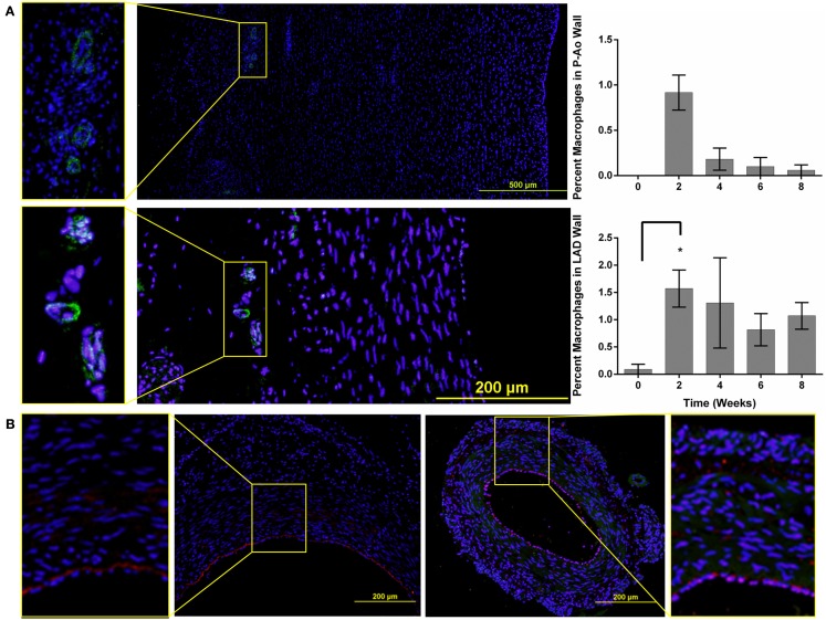
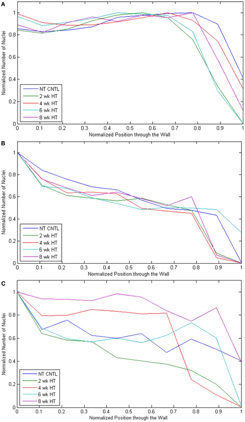
Methods: We used an aortic coarctation model of hypertension in the mini-pig to elucidate spatiotemporal changes in geometry and wall composition (including layer-specific thicknesses as well as presence of collagen, elastin, smooth muscle, endothelial, macrophage, and hematopoietic cells) in three different arterial beds, specifically aortic, cerebral, and coronary, and vasodilator function in two different arteriolar beds, the cerebral and coronary.
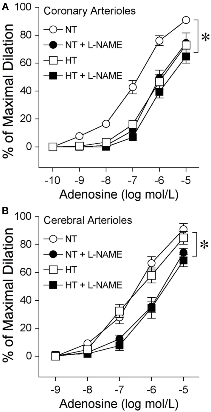
Results: Marked geometric and structural changes occurred in the thoracic aorta and left anterior descending coronary artery within 2 weeks of the establishment of hypertension and continued to increase over the 8-week study period. In contrast, no significant changes were observed in the middle cerebral arteries from the same animals. Consistent with these differential findings at the arterial level, we also found a diminished nitric oxide-mediated dilation to adenosine at 8 weeks of hypertension in coronary arterioles, but not cerebral arterioles.
Conclusion: These findings, coupled with the observation that temporal changes in wall constituents and the presence of macrophages differed significantly between the thoracic aorta and coronary arteries, confirm a strong differential progressive remodeling within different vascular beds. Taken together, these results suggest a spatiotemporal progression of vascular remodeling, beginning first in large elastic arteries and delayed in distal vessels.
Keywords: aorta; arterial remodeling; arteriolar function; collagen; elastin; neointima; vascular smooth muscle cell.
Figures


Similar articles
-
Upregulation of vascular arginase in hypertension decreases nitric oxide-mediated dilation of coronary arterioles.Hypertension. 2004 Dec;44(6):935-43. doi: 10.1161/01.HYP.0000146907.82869.f2. Epub 2004 Oct 18. Hypertension. 2004. PMID: 15492130
-
Computational simulations of hemodynamic changes within thoracic, coronary, and cerebral arteries following early wall remodeling in response to distal aortic coarctation.Biomech Model Mechanobiol. 2013 Jan;12(1):79-93. doi: 10.1007/s10237-012-0383-x. Epub 2012 Mar 14. Biomech Model Mechanobiol. 2013. PMID: 22415052 Free PMC article.
-
Regulation of coronary blood flow during exercise.Physiol Rev. 2008 Jul;88(3):1009-86. doi: 10.1152/physrev.00045.2006. Physiol Rev. 2008. PMID: 18626066 Review.
-
Functional and structural adaptations of coronary microvessels distal to a chronic coronary artery stenosis.Circ Res. 2008 Apr 11;102(7):795-803. doi: 10.1161/CIRCRESAHA.108.172528. Epub 2008 Feb 21. Circ Res. 2008. PMID: 18292598
-
Vascular gap junctions and implications for hypertension.Clin Exp Pharmacol Physiol. 2004 Oct;31(10):659-67. doi: 10.1111/j.1440-1681.2004.04071.x. Clin Exp Pharmacol Physiol. 2004. PMID: 15554905 Review.
Cited by
-
Hypertension: Causes and Consequences of Circadian Rhythms in Blood Pressure.Circ Res. 2024 Mar 15;134(6):810-832. doi: 10.1161/CIRCRESAHA.124.323515. Epub 2024 Mar 14. Circ Res. 2024. PMID: 38484034 Free PMC article. Review.
-
Exercise training in hypertension: Role of microRNAs.World J Cardiol. 2014 Aug 26;6(8):713-27. doi: 10.4330/wjc.v6.i8.713. World J Cardiol. 2014. PMID: 25228951 Free PMC article. Review.
-
Mechanobiological model of arterial growth and remodeling.Biomech Model Mechanobiol. 2018 Feb;17(1):87-101. doi: 10.1007/s10237-017-0946-y. Epub 2017 Aug 19. Biomech Model Mechanobiol. 2018. PMID: 28823079 Free PMC article.
-
Cerebral microhemorrhages: mechanisms, consequences, and prevention.Am J Physiol Heart Circ Physiol. 2017 Jun 1;312(6):H1128-H1143. doi: 10.1152/ajpheart.00780.2016. Epub 2017 Mar 17. Am J Physiol Heart Circ Physiol. 2017. PMID: 28314762 Free PMC article. Review.
-
A Systems Approach to Biomechanics, Mechanobiology, and Biotransport.J Biomech Eng. 2024 Apr 1;146(4):040801. doi: 10.1115/1.4064547. J Biomech Eng. 2024. PMID: 38270930 Free PMC article.
References
-
- Abcam (2010). Ihc Paraffin Staining Protocol. Cambridge: Abcam plc
-
- Armstead W. M. (1997). Role of nitric oxide, cyclic nucleotides, and the activation of ATP-sensitive K+ channels in the contribution of adenosine to hypoxia-induced pial artery dilation. J. Cereb. Blood Flow Metab. 17, 100–108 - PubMed
Grants and funding
LinkOut - more resources
Full Text Sources