. 2012 Nov 9:3:335.
doi: 10.3389/fimmu.2012.00335. eCollection 2012.

Human NK cell lytic granules and regulation of their exocytosis

Affiliations

Human NK cell lytic granules and regulation of their exocytosis

Konrad Krzewski et al. Front Immunol. .

Abstract

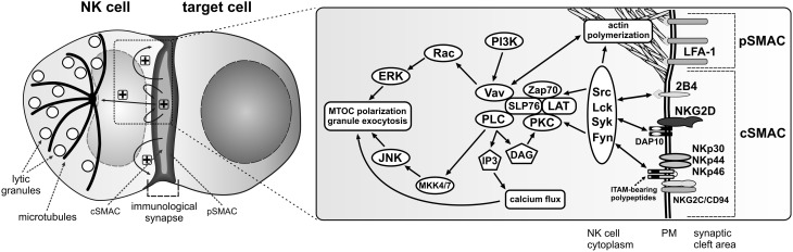
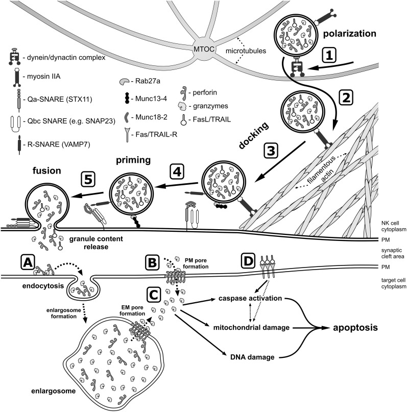
Natural killer (NK) cells form a subset of lymphocytes that play a key role in immuno-surveillance and host defense against cancer and viral infections. They recognize stressed cells through a variety of germline-encoded activating cell surface receptors and utilize their cytotoxic ability to eliminate abnormal cells. Killing of target cells is a complex, multi-stage process that concludes in the directed secretion of lytic granules, containing perforin and granzymes, at the immunological synapse. Upon delivery to a target cell, perforin mediates generation of pores in membranes of target cells, allowing granzymes to access target cell cytoplasm and induce apoptosis. Therefore, lytic granules of NK cells are indispensable for normal NK cell cytolytic function. Indeed, defects in lytic granule secretion lead or are related to serious and often fatal diseases, such as familial hemophagocytic lymphohistiocytosis (FHL) type 2-5 or Griscelli syndrome type 2. A number of reports highlight the role of several proteins involved in lytic granule release and NK cell-mediated killing of tumor cells. This review focuses on lytic granules of human NK cells and the advancements in understanding the mechanisms controlling their exocytosis.

Keywords: NK cells; cytotoxic lymphocytes; cytotoxicity; exocytosis; lysosomes; lytic granules.


Figures

Figure 1
Figure 1
Activation signals for lytic granule polarization in NK cells. The encounter between an NK cell and a susceptible target cell results in conjugation and formation of the activating immunological synapse. Adhesion molecules, such as LFA-1, segregate into the outer region of the synapse, referred to as the peripheral supramolecular activation cluster (pSMAC), while NK cell activating receptors localize into the central area of the synapse (cSMAC). Engagement of NK cell activating receptors by their ligands on the target cell (not shown) induces phosphorylation of membrane proximal signaling molecules and formation of a signalosome comprised of many signaling and adapter molecules at the cSMAC. Positive feedback loops are generated, causing signal amplification and sustained signaling (“+” symbols) that stimulates more robust actin polymerization at the synapse periphery and polarization of the MTOC and lytic granules to the immunological synapse, where the granules will be subsequently exocytosed. The diagrams represent only selected molecules. The drawings are not to scale.
Figure 2
Figure 2
A model of lytic granule exocytosis from human NK cell. In response to the engagement of NK cell activating receptors and initiation of signaling cascades (not depicted), the lytic granules move along the microtubules toward the MTOC in the dynein-dynactin complex-dependent manner (1). The MTOC and the granules then polarize toward the NK-target cell contact area, where granules switch from microtubules to the filamentous actin network at the immunological synapse (2) and navigate through the cortical filamentous actin meshwork as a result of the actin motor protein myosin IIA activity (3). This allows the lytic granules to get into close proximity of the plasma membrane (PM), and dock at the membrane due to activity of Rab27a and Rab27a-mediated recruitment of Munc13-4, as well as through the recognition of syntaxin 11 (STX11) and Munc18-2, possibly by the R-SNARE protein(s) present at the lytic granule membrane. The docked granules are then primed (4) by Munc13-4 in response to calcium flux (not shown), likely by the Munc13-4-mediated switch of STX11 to an open conformation (by removal of Munc18-2), and/or by Munc13-4 forming an initial bridge between the granule membrane and the plasma membrane. Finally, the granule-associated R-SNARE protein(s) (e.g., VAMP7) form a complex with Q-SNARE proteins present on the plasma membrane (e.g., STX11 and SNAP23) (5), which allows for the fusion of vesicles with the plasma membrane and release of the granule content into the synaptic cleft at the immunological synapse. There are two paradigms describing the entry of perforin and granzymes into target cells. The internalization model (A) assumes that perforin and granzymes bind to the target cell plasma membrane and are endocytosed into the early endosome-like enlargosome. Following internalization, perforin would mediate formation of pores in the enlargosome membrane (EM), allowing granzymes to leak into the cytosol of the target cell. According to the plasma membrane (PM) pore formation model (B), perforin oligomerizes in the plasma membrane, disrupting its integrity thereby permitting granzymes to enter from the synaptic cleft into the target cell. After gaining access into the cell cytosol (C), granzymes start processing their targets, leading to apoptosis through activation of caspases, induction of mitochondrial damage, and DNA fragmentation. In addition, FasL and TRAIL from the lytic granules bind to their receptors on the target cell surface (D) and initiate apoptosis.

Similar articles

Cited by

References

    1. Adrain C., Murphy B. M., Martin S. J. (2005). Molecular ordering of the caspase activation cascade initiated by the cytotoxic T lymphocyte/natural killer (CTL/NK) protease granzyme B. J. Biol. Chem. 280, 4663–4673 10.1074/jbc.M410915200 - DOI - PubMed
    1. Agerberth B., Charo J., Werr J., Olsson B., Idali F., Lindbom L., et al. (2000). The human antimicrobial and chemotactic peptides LL-37 and alpha-defensins are expressed by specific lymphocyte and monocyte populations. Blood 96, 3086–3093 - PubMed
    1. Aktas E., Kucuksezer U. C., Bilgic S., Erten G., Deniz G. (2009). Relationship between CD107a expression and cytotoxic activity. Cell. Immunol. 254, 149–154 10.1016/j.cellimm.2008.08.007 - DOI - PubMed
    1. Almeida C. R., Ashkenazi A., Shahaf G., Kaplan D., Davis D. M., Mehr R. (2011). Human NK cells differ more in their KIR2DL1-dependent thresholds for HLA-Cw6-mediated inhibition than in their maximal killing capacity. PLoS ONE 6:e24927 10.1371/journal.pone.0024927 - DOI - PMC - PubMed
    1. Almeida C. R., Davis D. M. (2006). Segregation of HLA-C from ICAM-1 at NK cell immune synapses is controlled by its cell surface density. J. Immunol. 177, 6904–6910 - PubMed

LinkOut - more resources