. 2012 Nov 12:3:251.
doi: 10.3389/fpls.2012.00251. eCollection 2012.

The putative K(+) channel subunit AtKCO3 forms stable dimers in Arabidopsis

Affiliations

The putative K(+) channel subunit AtKCO3 forms stable dimers in Arabidopsis

Alessandra Rocchetti et al. Front Plant Sci. .

Abstract

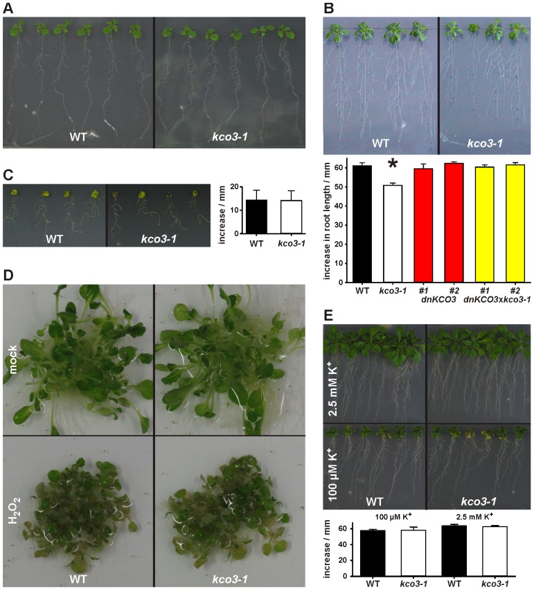
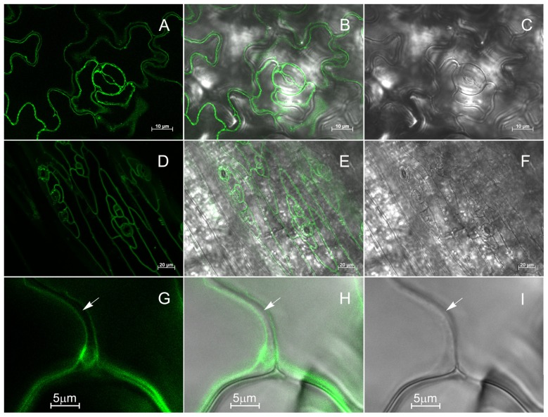
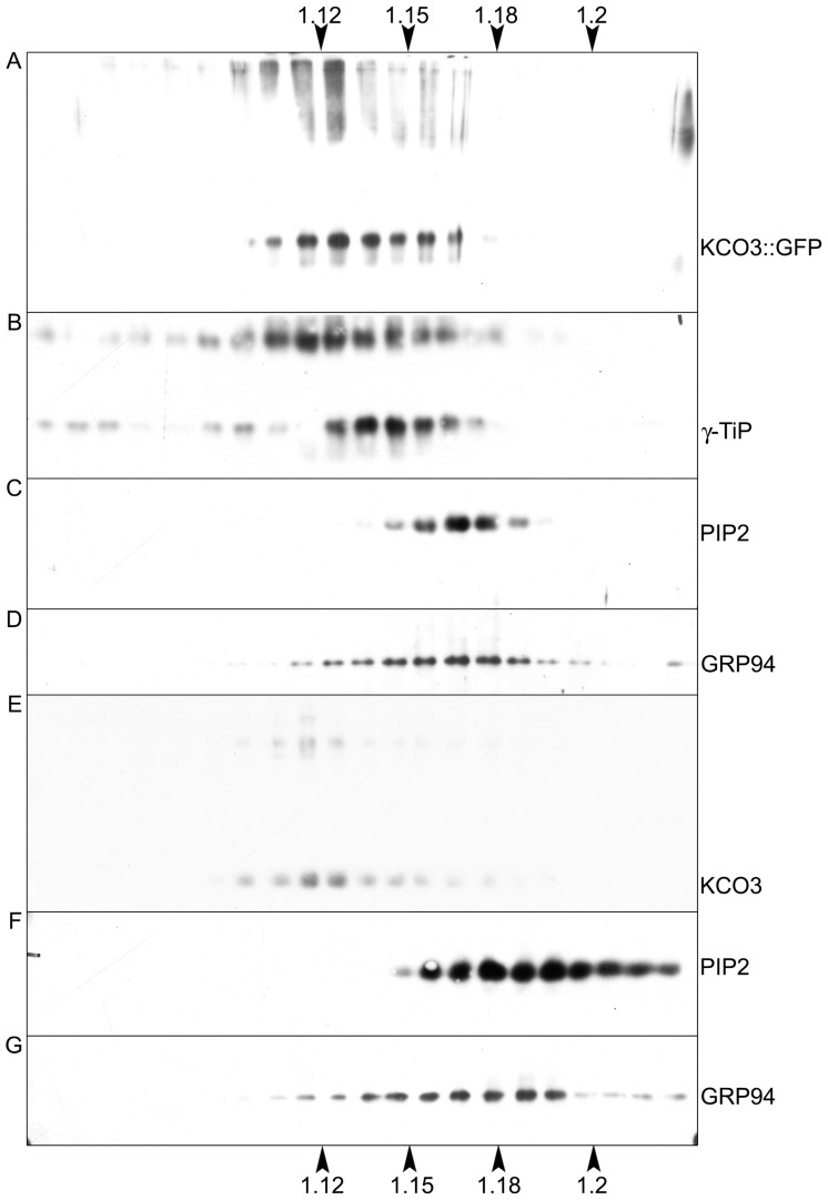
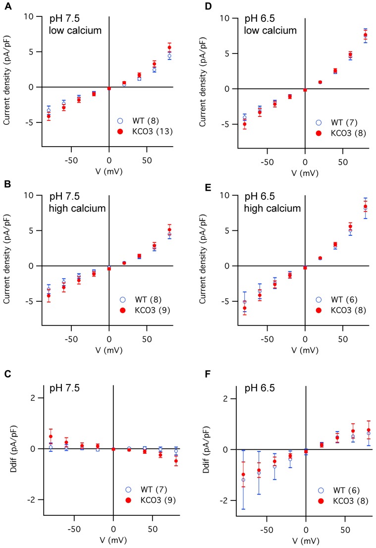
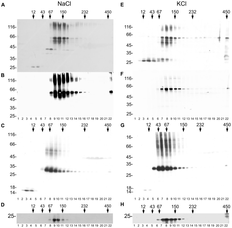
The permeation pore of K(+) channels is formed by four copies of the pore domain. AtKCO3 is the only putative voltage-independent K(+) channel subunit of Arabidopsis thaliana with a single pore domain. KCO3-like proteins recently emerged in evolution and, to date, have been found only in the genus Arabidopsis (A. thaliana and A. lyrata). We show that the absence of KCO3 does not cause marked changes in growth under various conditions. Only under osmotic stress we observed reduced root growth of the kco3-1 null-allele line. This phenotype was complemented by expressing a KCO3 mutant with an inactive pore, indicating that the function of KCO3 under osmotic stress does not depend on its direct ability to transport ions. Constitutively overexpressed AtKCO3 or AtKCO3::GFP are efficiently sorted to the tonoplast indicating that the protein is approved by the endoplasmic reticulum quality control. However, vacuoles isolated from transgenic plants do not have significant alterations in current density. Consistently, both AtKCO3 and AtKCO3::GFP are detected as homodimers upon velocity gradient centrifugation, an assembly state that would not allow for activity. We conclude that if AtKCO3 ever functions as a K(+) channel, active tetramers are held by particularly weak interactions, are formed only in unknown specific conditions and may require partner proteins.

Keywords: Arabidopsis; membrane proteins; potassium channels; protein assembly; tonoplast.


Figures

FIGURE 1
FIGURE 1
Genotyping of kco3-1 null-allele, dnKCO3 and kco3-1 × dnKCO3 mutant plants. (A) Schematic representation of the Salk_96038 T-DNA insertion line that contains two head to head T-DNA insertions at position 420 in the first exon of the KCO3 gene. (B) Schematic representation of dnKCO3 mutant, where, in the first exon of the KCO3 gene, a dominant negative mutation has been created by mutating the GFGD motif to GRGD. Additionally, a recognition site of the restriction enzyme SacII has been inserted. (C,D) PCR was performed on genomic DNA with the indicated primer sets specific for T-DNA amplification. Amplification was attained in case of kco3-1 and kco3-1 × dnKCO3 while the wild-type and dnKCO3 does not show any amplification product with the T-DNA primer sets. (E) PCR was performed with the indicated gene specific primer pair. Amplification can be observed in Col-0 wild-type, dnKCO3 and kco3-1 × dnKCO3. No amplification product is detectable in the homozygous T-DNA insertion line (kco3-1). (F) SacII restriction digestion of the PCR products obtained by amplification with the gene specific primer pair. PCR product from Col-0 wild-type was not digested. The amplification product from the dnKCO3 mutant was digested but some undigested product can also be seen. The PCR product from kco3-1 × dnKCO3 shows complete digestion.
FIGURE 2
FIGURE 2
Phenotypic analysis of kco3-1. (A) Growth on MS medium supplemented with 3% sucrose. The wild-type and the kco3-1 knock-out mutant do not show significant differences in shoot development and root length. Figure is representative of three independent experiments. (B) Osmotic stress. The figure is representative of three independent experiments. Lower panel: increase in root length after 15 days. The values shown are mean of six repeats ± SD (indicated by error bars). Data were analyzed using Student’s t-test. The values obtained for kco3-1 are significantly different from those of the WT control, dnKCO3 and kco3-1 × dnKCO3 plants (P < 0.001). (C) Salt stress. Growth of Col-0 and kco3-1 plants was severely hampered by the presence of NaCl. The figure is representative for three independent experiments. Right panel: increase in root length after 15 days. The values shown are mean of six repeats ± SD (indicated by error bars). Student’s t-test revealed that Col-0 and kco3-1 are not significantly different (P > 0.1). (D) Oxidative stress. After five days of H2O2 stress, Col-0 and kco3-1 plants showed symptoms such as bleaching of leaves and plant decay. The figure is representative for two independent experiments. (E) K+ deficient and K+ sufficient medium. Lower panel: increase in root length after 15 days was plotted as means ± SD (indicated by error bars) of six repeats. Student’s t-test did not reveal any significant difference between Col-0 and kco3-1 (P > 0.1).
FIGURE 3
FIGURE 3
Selection of Arabidopsis plants overexpressing KCO3::GFP and KCO3. (A) Equal amounts of leaf extracts (50 µg of total protein) from wild-type plants (Co) and independent transgenic Arabidopsis plants expressing KCO3::GFP (plants 1–11) were subjected to SDS-PAGE and western blot analysis with anti-GFP antiserum. The positions of the entire fusion protein and of free GFP are indicated. (B) Leaf extracts from one wild-type plant (lane 1) and transgenic plant n° 6 of panel A (lane 2) were analyzed by western blot using anti-KCO3 antibodies. The positions of monomers (arrowhead) and putative dimers (circle) are indicated. (C) Equal amounts (50 µg of total protein) of leaf extracts from wild-type (Co) or independent transgenic Arabidopsis plants overexpressing KCO3 (plants 2–7) were analyzed by western blot with anti-KCO3 antiserum. The positions of KCO3 (arrow) and an unspecific, cross-reacting polypeptide (asterisk) are indicated. In each panel, numbers on the left indicate the position and size (in kDa) of molecular mass markers.
FIGURE 4
FIGURE 4
KCO3::GFP is located at the tonoplast. Leaf epidermis (A–C, G–I) or hypocotyl (D–F) from KCO3::GFP transgenic plant were analyzed by epifluorescence microscopy. (A,D,G): GFP fluorescence; (C,F,I): brightfield; (B,E,H): Merge of fluorescence and brightfield. The arrow indicates the cell surface.
FIGURE 5
FIGURE 5
KCO3::GFP and KCO3 co-fractionate with a tonoplast marker in isopycnic gradients. Young leaves from transgenic Arabidopsis plants expressing KCO3::GFP (A–D) or KCO3 (E–G) were homogenized in the presence of sucrose and the absence of detergent. Microsomes were pelleted and further separated by centrifugation on isopycnic sucrose gradient. Proteins in each fraction (1/10 of fraction volume) were analyzed by SDS-PAGE and western blot with antibodies against KCO3 (to detect KCO3::GFP or KCO3), γTIP (tonoplast marker), PIP2 (plasma membrane marker) or endoplasmin (GRP94, endoplasmic reticulum marker), as indicated at the right of each panel. Top of each gradient is at left; numbers on top or bottom of the figure indicate fraction density (g/ml).
FIGURE 6
FIGURE 6
Current density versus voltage characteristics recorded in different experimental conditions from vacuoles of Arabidopsis wild-type and KCO3-overexpressing plants. (A–C) Current density–voltage relationships recorded in the whole-vacuole configuration (see Figure A1) from vacuoles of Arabidopsis wild-type (open blue symbols) and KCO3-overexpressing plants (filled red symbols). Bath solutions of pH 7.5 contained either low calcium (A) or high calcium (B). The current density difference (Ddif) between high and low calcium conditions was calculated for individual vacuoles and averaged (C). (D,F) As in (A–C), but in bath solutions of pH 6.5, in low calcium (D), in high calcium (E), difference (F; Ddif). In each panel, the number of WT or KCO3 vacuoles analyzed is given in brackets.
FIGURE 7
FIGURE 7
KCO3::GFP and KCO3 are dimers. Leaves from KCO3::GFP (A,B,D–F,H) or KCO3 (C,G) transgenic Arabidopsis plants were homogenized in the presence of non-ionic detergent and either 200 mM NaCl (A–D) or 40 mM KCl (E–H). The homogenates were then subjected to sedimentation velocity centrifugation on a continuous 5–25% (w/v) sucrose gradient. One-tenth of the total volume of each gradient fraction were analyzed by SDS-PAGE and western blot using anti-GFP antiserum to detect KCO3::GFP (A,E), anti-KCO3 antiserum to detect KCO3::GFP (B,F) or KCO3 (C,G), anti-γTIP (D,H). Top of each gradient is at left. Numbers on top indicate the position along the gradient and the molecular mass (in kDa) of sedimentation markers. Numbers at left indicate the positions of SDS-PAGE molecular mass markers.
Figure A1
Figure A1
Current traces recorded in whole-vacuole mode in vacuoles isolated from Arabidopsis mesophyll cells. (A) Current traces recorded 2 min after achieving the whole-vacuole configuration in response to voltage steps from +80 to -80 mV, step -20 mV. The holding and tail voltages were 0 and -50 mV, respectively. The vacuole had a capacitance of 34 pF and derived from an Arabidopsis wild-type plant. (B) Current traces recorded from the same vacuole as in A, 20 min after the break-in.

Similar articles

Cited by

References

    1. Allen G. J., Amtmann A., Sanders D. (1998). Calcium-dependent and calcium-independent K+ mobilization channels in Vicia faba guard cell vacuoles. J. Exp. Bot. 49 305–318
    1. Allen G. J., Sanders D. (1996). Control of ionic currents in guard cell vacuoles by cytosolic and luminal calcium. Plant J. 10 1055–1069 - PubMed
    1. Dadacz-Narloch B., Beyhl D., Larisch C., Lopez-Sanjurjo E. J., Reski R., Kuchitsu K., et al. (2011). A novel calcium binding site in the slow vacuolar cation channel TPC1 senses luminal calcium levels. Plant Cell 23 2696–2707 - PMC - PubMed
    1. Devereux J., Haeberli P., Smithies O. (1984). A comprehensive set of sequence analysis programs for the VAX. Nucleic Acids Res. 12 387–395 - PMC - PubMed
    1. Dreyer I., Antunes S., Hoshi T., Müller-Röber B., Palme K., Pongs O., et al. (1997). Plant K+ channel α-subunits assemble indiscriminately. Biophys. J. 72 2143–2150 - PMC - PubMed

LinkOut - more resources