

Expression of UDP-glucuronosyltransferase 1A, nuclear factor erythroid-E2-related factor 2 and Kelch-like ECH-associated protein 1 in colonic mucosa, adenoma and adenocarcinoma tissue
- PMID: 23162624
- PMCID: PMC3499601
- DOI: 10.3892/ol.2012.850
Expression of UDP-glucuronosyltransferase 1A, nuclear factor erythroid-E2-related factor 2 and Kelch-like ECH-associated protein 1 in colonic mucosa, adenoma and adenocarcinoma tissue
Abstract
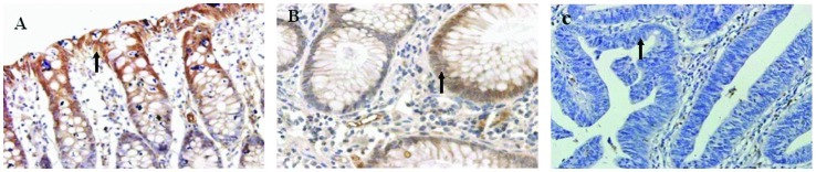
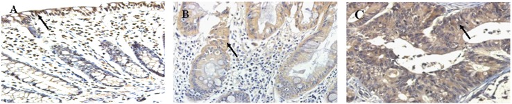
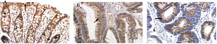
We investigated the expression of UDP-glucuronosyltransferase 1A (UGT1A), nuclear factor erythroid-E2-related factor 2 (Nrf2) and Kelch-like ECH-associated protein 1 (Keap1) in normal colonic mucosa, adenoma tissue and adenocarcinoma tissue, to analyze the correlation between their expression and the clinicopathological features of adenocarcinoma and their roles in colonic carcinogenesis. Using immunohistochemical analysis, we investigated the expression of UGT1A, Nrf2 and Keap1 in normal colonic mucosa (n=24), adenoma tissue (n=30) and adenocarcinoma tissue (n=77). We found that the positive rate of UGT1A was 83.3% in normal colonic mucosa, 80.0% in adenoma tissue and significantly lower, 53.2%, in adenocarcinoma tissue (normal colonic vs. adenoma tissue, P=0.071; normal colonic vs. adenocarcinoma tissue, P=0.023; adenoma vs. adenocarcinoma tissue, P=0.019). The expression levels of Nrf2 were high in adenoma and adenocarcinoma tissues, with positive rates of 70.0 and 87.0%, respectively, but were significantly lower in normal colonic tissue, with a positive rate of 41.7% (normal colonic vs. adenoma tissue, P=0.000; normal colonic vs. adenocarcinoma tissue, P=0.000; adenoma vs. adenocarcinoma tissue, P=0.000). The positive rate of Keap1 was 54.2% in normal mucosa, 70.0% in adenoma tissue and 61.0% in adenocarcinoma tissue (normal colonic mucosa vs. adenoma tissue, P=0.200; normal colonic vs. adenocarcinoma tissue, P=0.040; adenoma vs. adenocarcinoma, P=0.002). In addition, there was no correlation between the expression of Nrf2/Keap1 in adenoma and adenocarcinoma tissues (r=0.067, P=0.723; r=0.042, P=0.715, respectively). The results suggest that decreased expression of UGT1A and the dysregulation of the Nrf2/Keap1 system may be related to colonic carcinogenesis.
Figures
Similar articles
-
[The UGT1A, Nrf2 and Keap1 protein expression and significance in colon tumor].Zhonghua Nei Ke Za Zhi. 2012 Jul;51(7):531-5. Zhonghua Nei Ke Za Zhi. 2012. PMID: 22943825 Chinese.
-
[Nrf2 and Keap1 Abnormalities in 104 Lung Adenocarcinoma Cases and Association with Clinicopathologic Features].Zhongguo Fei Ai Za Zhi. 2018 Mar 20;21(3):241-250. doi: 10.3779/j.issn.1009-3419.2018.03.04. Zhongguo Fei Ai Za Zhi. 2018. PMID: 29587953 Free PMC article. Chinese.
-
Immunohistochemical study of the Nrf2 pathway in colorectal cancer: Nrf2 expression is closely correlated to Keap1 in the tumor and Bach1 in the normal tissue.Appl Immunohistochem Mol Morphol. 2013 Dec;21(6):511-7. doi: 10.1097/PAI.0b013e318282ac20. Appl Immunohistochem Mol Morphol. 2013. PMID: 23455180
-
Emerging Substrate Proteins of Kelch-like ECH Associated Protein 1 (Keap1) and Potential Challenges for the Development of Small-Molecule Inhibitors of the Keap1-Nuclear Factor Erythroid 2-Related Factor 2 (Nrf2) Protein-Protein Interaction.J Med Chem. 2020 Aug 13;63(15):7986-8002. doi: 10.1021/acs.jmedchem.9b01865. Epub 2020 Apr 8. J Med Chem. 2020. PMID: 32233486 Review.
-
Nuclear factor (erythroid-derived 2)-like 2 (NRF2) drug discovery: Biochemical toolbox to develop NRF2 activators by reversible binding of Kelch-like ECH-associated protein 1 (KEAP1).Arch Biochem Biophys. 2017 Oct 1;631:31-41. doi: 10.1016/j.abb.2017.08.003. Epub 2017 Aug 8. Arch Biochem Biophys. 2017. PMID: 28801166 Review.
Cited by
-
Species differences in drug glucuronidation: Humanized UDP-glucuronosyltransferase 1 mice and their application for predicting drug glucuronidation and drug-induced toxicity in humans.Drug Metab Pharmacokinet. 2018 Feb;33(1):9-16. doi: 10.1016/j.dmpk.2017.10.002. Epub 2017 Oct 7. Drug Metab Pharmacokinet. 2018. PMID: 29079228 Free PMC article. Review.
-
Research progress on chemopreventive effects of phytochemicals on colorectal cancer and their mechanisms.World J Gastroenterol. 2016 Aug 21;22(31):7058-68. doi: 10.3748/wjg.v22.i31.7058. World J Gastroenterol. 2016. PMID: 27610016 Free PMC article. Review.
-
Sulforaphane suppresses carcinogenesis of colorectal cancer through the ERK/Nrf2‑UDP glucuronosyltransferase 1A metabolic axis activation.Oncol Rep. 2020 Apr;43(4):1067-1080. doi: 10.3892/or.2020.7495. Epub 2020 Feb 10. Oncol Rep. 2020. PMID: 32323779 Free PMC article.
-
Pathways Related to Colon Inflammation Are Associated with Colorectal Carcinoma: A Transcriptome- and Methylome-Wide Study.Cancers (Basel). 2023 May 26;15(11):2921. doi: 10.3390/cancers15112921. Cancers (Basel). 2023. PMID: 37296884 Free PMC article.
-
Sulforaphane-induced epigenetic regulation of Nrf2 expression by DNA methyltransferase in human Caco-2 cells.Oncol Lett. 2019 Sep;18(3):2639-2647. doi: 10.3892/ol.2019.10569. Epub 2019 Jul 5. Oncol Lett. 2019. PMID: 31452747 Free PMC article.
References
-
- Malfatti MA, Felton JS. N-glucuronidation of 2-amino-1-methyl-6-phenylimidazo[4,5-b]pyridine (PhIP) and N-hydroxy-PhIP by specific human UDP-glucuronosyltransferases. Carcinogenesis. 2001;22:1087–1093. - PubMed
-
- Giuliani L, Gazzanga P, Caporuscio F, et al. Can down-regulation of UDP-glucuronosyltransferases in the urinary bladder tissue impact the risk of chemical cacinogenesis? Int J Cancer. 2001;91:141–143. - PubMed
-
- McDonnell WM, Hitomi E, Askari FK. Identification of bilirubin UDP-GTs in the human alimentary tract in accordance with the gut as a putative metabolic organ. Biochem Pharmacol. 1996;51:483–488. - PubMed
LinkOut - more resources
Full Text Sources
Research Materials