. 2012 Oct;38(5):489-98.
doi: 10.1007/s00068-012-0229-3. Epub 2012 Sep 30.

Rationales for the Bernese approaches in acetabular surgery

Affiliations

Rationales for the Bernese approaches in acetabular surgery

M J B Keel et al. Eur J Trauma Emerg Surg. 2012 Oct.

Abstract

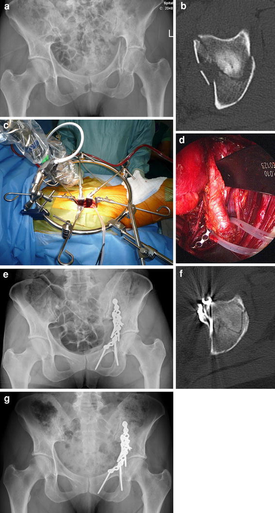
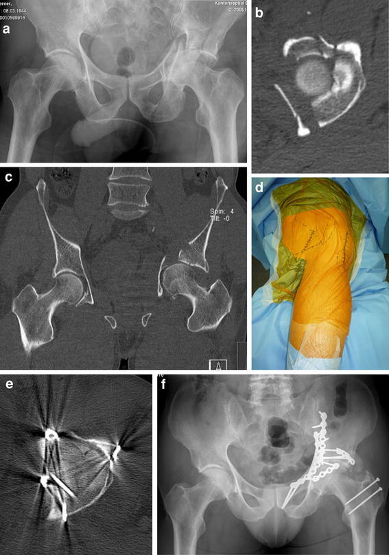
Purpose: To present two new approaches to acetabular surgery that were established in Berne, and which aim at enhanced visualization and anatomical reconstruction of acetabular fractures.

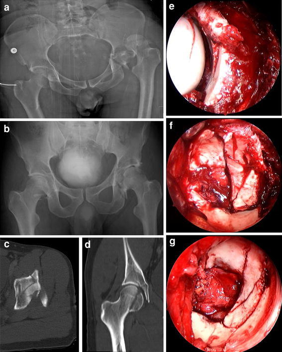
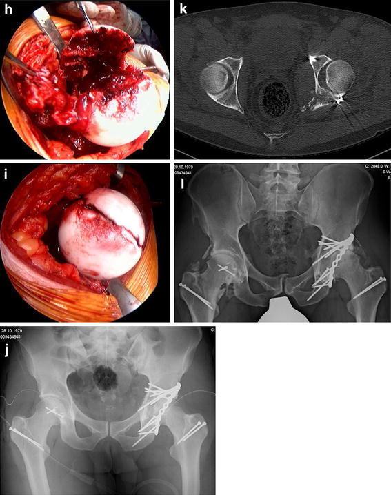
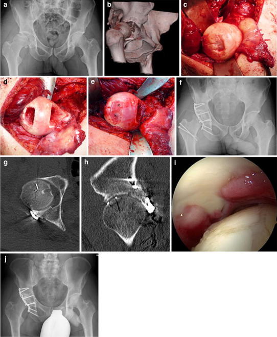
Method: The trochanteric flip osteotomy allows for surgical hip dislocation, and was introduced as a posterior approach for acetabular fracture management involving the posterior column and wall. For acetabular fractures predominantly involving the anterior column and the quadrilateral plate, the Pararectus approach is described.

Results: Full exposure of the hip joint, as provided by the trochanteric flip osteotomy, facilitates anatomical reduction of acetabular or femoral head fractures and safe positioning of the anterior column screw in transverse or T-shaped fractures. Additionally, the approach enables osteochondral transplantation as a salvage procedure for severe chondral femoral head damage and osteoplasty of an associated inadequate offset at the femoral head-neck junction. The Pararectus approach allows anatomical restoration with minimal access morbidity, and combines advantages of the ilioinguinal and modified Stoppa approaches.

Conclusions: Utilization of the trochanteric flip osteotomy eases visualization of the superior aspect of the acetabulum, and enables the evaluation and treatment of chondral lesions of the femoral head or acetabulum and labral tears. Displaced fractures of the anterior column with a medialized quadrilateral plate can be addressed successfully through the Pararectus approach, in which surgical access is associated with minimal morbidity. However, long-term results following the two presented Bernese approaches are needed to confirm that in the treatment of complex acetabular fractures the rate of poor results in almost one-third of all cases (as currently yielded using traditional approaches) might be reduced by the utilization of the presented novel approaches.


Figures

Fig. 1
Fig. 1
a Anteroposterior radiograph of the pelvis of a 30-year-old patient who sustained bilateral posterior hip dislocations due to a work accident before closed reduction. b Anteroposterior radiograph of the pelvis after closed reduction. c Axial CT scan shows a transverse acetabular fracture with comminuted posterior wall fracture of the left hip. d Coronal CT scan demonstrates a femoral head fracture (Pipkin type I) on the right side. e Intraoperative view of the left acetabulum through the posterior approach shows marginal impaction and comminuted posterior wall fracture. f Trochanteric flip osteotomy and surgical hip dislocation allows complete exposure of the transverse fracture with comminuted posterior wall fracture. g Exposure of the acetabulum after anatomic reduction and internal fixation. h Intraoperative view of the right femoral head before anatomic reduction and fixation after surgical hip dislocation. i Intraoperative view of the right femoral head after anatomic reduction and fixation. j Postoperative anteroposterior radiograph of the pelvis demonstrates anatomic reconstructions of the Pipkin I fracture on the right side and of the acetabular fracture on the left side. k Postoperative CT scan demonstrates anatomic reconstructions of the Pipkin I fracture on the right side and of the acetabular fracture on the left side. l Anteroposterior radiograph of the pelvis 3 years after surgery shows bilateral ectopic ossifications (Brooker grade 2) and moderate signs of posttraumatic osteoarthritis of the left hip of the patient with an excellent clinical result
Fig. 1
Fig. 1
a Anteroposterior radiograph of the pelvis of a 30-year-old patient who sustained bilateral posterior hip dislocations due to a work accident before closed reduction. b Anteroposterior radiograph of the pelvis after closed reduction. c Axial CT scan shows a transverse acetabular fracture with comminuted posterior wall fracture of the left hip. d Coronal CT scan demonstrates a femoral head fracture (Pipkin type I) on the right side. e Intraoperative view of the left acetabulum through the posterior approach shows marginal impaction and comminuted posterior wall fracture. f Trochanteric flip osteotomy and surgical hip dislocation allows complete exposure of the transverse fracture with comminuted posterior wall fracture. g Exposure of the acetabulum after anatomic reduction and internal fixation. h Intraoperative view of the right femoral head before anatomic reduction and fixation after surgical hip dislocation. i Intraoperative view of the right femoral head after anatomic reduction and fixation. j Postoperative anteroposterior radiograph of the pelvis demonstrates anatomic reconstructions of the Pipkin I fracture on the right side and of the acetabular fracture on the left side. k Postoperative CT scan demonstrates anatomic reconstructions of the Pipkin I fracture on the right side and of the acetabular fracture on the left side. l Anteroposterior radiograph of the pelvis 3 years after surgery shows bilateral ectopic ossifications (Brooker grade 2) and moderate signs of posttraumatic osteoarthritis of the left hip of the patient with an excellent clinical result
Fig. 2
Fig. 2
a Anteroposterior radiograph of the pelvis of a 44-year-old patient one week after a car accident demonstrates a partially dislocated femoral head on the right side, intraarticular bone fragments, and a comminuted posterior wall acetabular fracture. b Three-dimensional CT reconstruction demonstrates a partially dislocated femoral head on the right side, intraarticular bone fragments, and a comminuted posterior wall acetabular fracture. c Intraoperative view of the right femoral head shows the osteochondral defect within the weight-bearing area and the ruptured hip capsule with an intact retinaculum. d View of the femoral head after resection of the damaged femoral area and after the harvesting of an osteochondral shell autograft within the femoral head–neck junction. e Superior view after reconstruction of the articular surface of the femoral head using the graft and fixation with two 1.5 mm screws. f Postoperative anteroposterior radiograph of the pelvis demonstrates anatomic reconstruction of the posterior wall fracture and femoral head damage with osteochondral transplantation. g Axial CT scan demonstrates anatomic reconstruction of the posterior wall fracture and femoral head damage with osteochondral transplantation. h Sagittal CT reconstruction seven months postoperatively shows complete integration of the graft and the two slightly prominent screws in the femoral head, as well as small cyst formation in the dome as a sign of early osteoarthritis. i Arthroscopic view of the acetabular and femoral head cartilages shows moderate chondropathy of the femoral head during hip arthroscopy eight months after trauma from partial removal of the femoral head screws and removal of the trochanter screws. j Anteroposterior radiograph of the pelvis 1 year after surgical hip dislocation
Fig. 3
Fig. 3
a Intraoperative view after a “pararectal” skin incision that starts cranially at the border between the lateral and middle thirds of the line connecting the umbilicus and the anterior superior iliac spine (ASIS), and is directed to the border between the middle and medial thirds of the line connecting the ASIS with the symphysis. After incision of the anterior rectus sheath, the rectus abdominis muscle and the transversalis fascia are visualized. b Intraoperative view of the right hemipelvis showing the second, third, and fourth window developed by the Pararectus approach with the external iliac vessels, the vas deferens, and the inferior epigastric vessels indicating the borders of particular windows. c Lateral retraction of the external iliac vessels provides access to the fifth window, with visualization of the quadrilateral plate and the obturator nerve
Fig. 4
Fig. 4
a Anteroposterior radiograph of the pelvis of a 56-year-old female patient after a bicycle accident demonstrates a both-column acetabular fracture with a medialized quadrilateral plate. b Axial CT scan of the pelvis demonstrates a both-column acetabular fracture with a medialized quadrilateral plate. c Intraoperative view of the Pararectus approach and operative setting using the retractor system (Synframe) and endoscope to allow a less invasive approach. d Endoscopic view of the obturator nerve (left), reconstruction plates on the pelvic brim and quadrilateral plate, and the mobilized external iliac vessels. e Postoperative anteroposterior radiograph of the pelvis demonstrates anatomic reconstruction of the both column fracture. f Axial CT scan demonstrates anatomic reconstruction of the both-column fracture. g Anteroposterior radiograph of the pelvis 2 years after surgery without any signs of osteoarthritis. The patient showed an excellent clinical result after only 6 months postoperatively and is now working as a yoga teacher
Fig. 5
Fig. 5
a Anteroposterior radiograph of the pelvis of a 67-year-old patient following a bicycle accident that caused a both-column acetabular fracture with central femoral head dislocation, a medialized quadrilateral plate, and dome impression. b Axial CT scan of the pelvis demonstrates a both-column acetabular fracture with central femoral head dislocation, a medialized quadrilateral plate, and dome impression. c Coronal CT scan of the pelvis demonstrates a both-column acetabular fracture with central femoral head dislocation, a medialized quadrilateral plate, and dome impression. d Intraoperative view of the floppy semilateral position that allows simultaneous anterior access by the Pararectus approach and posterior access by the trochanteric flip osteotomy and surgical hip dislocation. e Postoperative axial CT scan showing anatomic reconstruction of the left hip. f Anteroposterior radiograph of the pelvis 1 year after surgery, with some ectopic ossifications present posteriorly (Brooker grade 2)

Similar articles

Cited by

References

    1. Giannoudis PV, Tzioupis C, Papathanassopoulos A, Obakponovwe O, Roberts C. Articular step-off and risk of post-traumatic osteoarthritis. Evidence today. Injury. 2010;41:986–995. doi: 10.1016/j.injury.2010.08.003. - DOI - PubMed
    1. Giannoudis PV, Grotz MR, Papakostidis C, Dinopoulos H. Operative treatment of displaced fractures of the acetabulum. A meta-analysis. J Bone Joint Surg (Br) 2005;87-B:2–9. - PubMed
    1. Letournel E. Acetabulum fractures: classification and management. Clin Orthop. 1980;151:81–106. - PubMed
    1. Briffa N, Pearce R, Hill AM, Bircher M. Outcomes of acetabular fracture fixation with ten years’ follow-up. J Bone Joint Surg (Br) 2011;93-B:229–236. doi: 10.1302/0301-620X.93B2.24056. - DOI - PubMed
    1. Anglen JO, Burd TA, Hendricks KJ, Harrision P. The “gull sign”: a harbinger of failure for internal fixation of geriatric acetabular fractures. J Orthop Trauma. 2003;17:625–34. - PubMed