

Surgical management of osteoporotic pelvic fractures: a new challenge
- PMID: 23162670
- PMCID: PMC3495273
- DOI: 10.1007/s00068-012-0224-8
Surgical management of osteoporotic pelvic fractures: a new challenge
Abstract
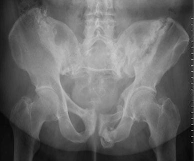
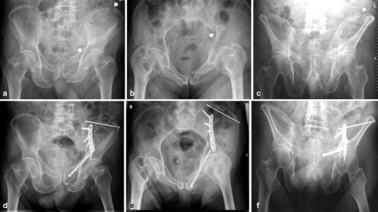
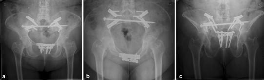
The number and variety of osteoporotic fractures of the pelvis are rapidly growing around the world. Such fractures are the result of low-impact trauma. The patients have no signs of hemodynamic instability and do not require urgent stabilization. The clinical picture is dominated by immobilizing pain in the pelvic region. Fractures may be located in both the ventral and the dorsal pelvic ring. The current well-established classification of pelvic ring lesions in younger adults does not fully reflect the criteria for osteoporotic and insufficiency fractures of the pelvic ring. Most osteoporotic fractures are minimally displaced and do not require surgical therapy. However, in some patients, an insidious progress of bone damage leads to complex displacement and instability. Therefore, vertical sacral ala fractures, fracture dislocations of the sacroiliac joint, and spinopelvic dissociations are best treated with operative stabilization. Angular stable bridge plating, the insertion of a transsacral positioning bar, and iliolumbar fixation are operative techniques that have been adapted to the low bone mineral density of the pelvic ring and the high forces acting on it.
Figures
Similar articles
-
When and How to Operate Fragility Fractures of the Pelvis?Indian J Orthop. 2019 Jan-Feb;53(1):128-137. doi: 10.4103/ortho.IJOrtho_631_17. Indian J Orthop. 2019. PMID: 30905993 Free PMC article.
-
Do Transsacral-transiliac Screws Across Uninjured Sacroiliac Joints Affect Pain and Functional Outcomes in Trauma Patients?Clin Orthop Relat Res. 2016 Jun;474(6):1417-21. doi: 10.1007/s11999-015-4596-z. Clin Orthop Relat Res. 2016. PMID: 26472585 Free PMC article.
-
[Osteoporotic fractures of the pelvic ring].Z Orthop Unfall. 2012 Jun;150(3):e107-18; quiz e119-20. doi: 10.1055/s-0032-1314948. Epub 2012 Jun 21. Z Orthop Unfall. 2012. PMID: 22723074 German.
-
Minimal Invasive Surgical Treatment of Fragility Fractures of the Pelvis.Chirurgia (Bucur). 2017 Sept-Oct;112(5):524-537. doi: 10.21614/chirurgia.112.5.524. Chirurgia (Bucur). 2017. PMID: 29088552 Review.
-
Osteosynthesis in sacral fracture and lumbosacral dislocation.Orthop Traumatol Surg Res. 2016 Feb;102(1 Suppl):S45-57. doi: 10.1016/j.otsr.2015.12.002. Epub 2016 Jan 22. Orthop Traumatol Surg Res. 2016. PMID: 26810715 Review.
Cited by
-
[Selection of access and positioning for operative treatment of pelvic injuries. Decision-making strategies].Unfallchirurg. 2013 Mar;116(3):227-37. doi: 10.1007/s00113-012-2331-8. Unfallchirurg. 2013. PMID: 23478900 German.
-
Analysis of the Spinopelvic Parameters in Patients with Fragility Fractures of the Pelvis.J Clin Med. 2023 Jul 2;12(13):4445. doi: 10.3390/jcm12134445. J Clin Med. 2023. PMID: 37445480 Free PMC article.
-
Pelvic and acetabular fractures.Eur J Trauma Emerg Surg. 2012 Oct;38(5):487-8. doi: 10.1007/s00068-012-0226-6. Epub 2012 Oct 16. Eur J Trauma Emerg Surg. 2012. PMID: 26816251 No abstract available.
-
Short-term follow-up of anterior and posterior both column fractures of acetabulum managed through both column plating.Eur J Orthop Surg Traumatol. 2019 Apr;29(3):605-610. doi: 10.1007/s00590-018-2331-7. Epub 2018 Oct 30. Eur J Orthop Surg Traumatol. 2019. PMID: 30377823
-
When and How to Operate Fragility Fractures of the Pelvis?Indian J Orthop. 2019 Jan-Feb;53(1):128-137. doi: 10.4103/ortho.IJOrtho_631_17. Indian J Orthop. 2019. PMID: 30905993 Free PMC article.
References
-
- Tile M, Hear T, Vrahas M. Biomechanics of the pelvic ring. In: Tile M, Helfet DL, Kellam JF, editors. Fractures of the pelvis and acetabulum. 3. Philadelphia: Lippincott Williams & Wilkins; 2003. pp. 32–45.
-
- Huber-Wagner S, Lefering R, Qvick LM, Körner M, Kay MV, Pfeifer KJ, Reiser M, Mutschler W, Kanz KG, Working Group on Polytrauma of the German Trauma Society Effect of whole-body CT during trauma resuscitation on survival: a retrospective, multicentre study. Lancet. 2009;373:1455–1461. doi: 10.1016/S0140-6736(09)60232-4. - DOI - PubMed
LinkOut - more resources
Full Text Sources
