

GENE EXPRESSION AND COLLAGEN FIBER MICROMECHANICAL INTERACTIONS OF THE SEMILUNAR HEART VALVE INTERSTITIAL CELL
- PMID: 23162672
- PMCID: PMC3498494
- DOI: 10.1007/s12195-012-0230-2
GENE EXPRESSION AND COLLAGEN FIBER MICROMECHANICAL INTERACTIONS OF THE SEMILUNAR HEART VALVE INTERSTITIAL CELL
Abstract
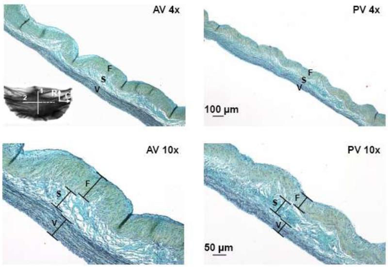
The semilunar (aortic and pulmonary) heart valves function under dramatically different hemodynamic environments, and have been shown to exhibit differences in mechanical properties, extracellular matrix (ECM) structure, and valve interstitial cell (VIC) biosynthetic activity. However, the relationship between VIC function and the unique micromechanical environment in each semilunar heart valve remains unclear. In the present study, we quantitatively compared porcine semilunar mRNA expression of primary ECM constituents, and layer- and valve-specific VIC-collagen mechanical interactions under increasing transvalvular pressure (TVP). Results indicated that the aortic valve (AV) had a higher fibrillar collagen mRNA expression level compared to the pulmonary valve (PV). We further noted that VICs exhibited larger deformations with increasing TVP in the collagen rich fibrosa layer, with substantially smaller changes in the spongiosa and ventricularis layers. While the VIC-collagen micro-mechanical coupling varied considerably between the semilunar valves, we observed that the VIC deformations in the fibrosa layer were similar at each valve's respective peak TVP. This result suggests that each semilunar heart valve's collagen fiber microstructure is organized to induce a consistent VIC deformation under its respective diastolic TVP. Collectively, our results are consistent with higher collagen biosynthetic demands for the AV compared to the PV, and that the valvular collagen microenvironment may play a significant role in regulating VIC function.
Figures


Similar articles
-
On intrinsic stress fiber contractile forces in semilunar heart valve interstitial cells using a continuum mixture model.J Mech Behav Biomed Mater. 2016 Feb;54:244-58. doi: 10.1016/j.jmbbm.2015.09.027. Epub 2015 Nov 11. J Mech Behav Biomed Mater. 2016. PMID: 26476967 Free PMC article.
-
Correlation between heart valve interstitial cell stiffness and transvalvular pressure: implications for collagen biosynthesis.Am J Physiol Heart Circ Physiol. 2006 Jan;290(1):H224-31. doi: 10.1152/ajpheart.00521.2005. Epub 2005 Aug 26. Am J Physiol Heart Circ Physiol. 2006. PMID: 16126816
-
In-situ deformation of the aortic valve interstitial cell nucleus under diastolic loading.J Biomech Eng. 2007 Dec;129(6):880-89. doi: 10.1115/1.2801670. J Biomech Eng. 2007. PMID: 18067392
-
On the Functional Role of Valve Interstitial Cell Stress Fibers: A Continuum Modeling Approach.J Biomech Eng. 2017 Feb 1;139(2):0210071-02100713. doi: 10.1115/1.4035557. J Biomech Eng. 2017. PMID: 28024085 Free PMC article. Review.
-
Heart valve structure and function in development and disease.Annu Rev Physiol. 2011;73:29-46. doi: 10.1146/annurev-physiol-012110-142145. Annu Rev Physiol. 2011. PMID: 20809794 Free PMC article. Review.
Cited by
-
Viability of Partial Heart Transplant Grafts During Prolonged Cold Preservation Suggests That Longer Donor Cold Chain Logistics May Be Feasible.Pediatr Transplant. 2025 May;29(3):e70063. doi: 10.1111/petr.70063. Pediatr Transplant. 2025. PMID: 40098581
-
Regulation of valve interstitial cell homeostasis by mechanical deformation: implications for heart valve disease and surgical repair.J R Soc Interface. 2017 Oct;14(135):20170580. doi: 10.1098/rsif.2017.0580. J R Soc Interface. 2017. PMID: 29046338 Free PMC article.
-
Learning-enhanced 3D fiber orientation mapping in thick cardiac tissues.Biomed Opt Express. 2025 Jul 22;16(8):3315-3336. doi: 10.1364/BOE.563643. eCollection 2025 Aug 1. Biomed Opt Express. 2025. PMID: 40809973 Free PMC article.
-
On the in vivo function of the mitral heart valve leaflet: insights into tissue-interstitial cell biomechanical coupling.Biomech Model Mechanobiol. 2017 Oct;16(5):1613-1632. doi: 10.1007/s10237-017-0908-4. Epub 2017 Apr 20. Biomech Model Mechanobiol. 2017. PMID: 28429161 Free PMC article.
-
Fibroblast growth factor 2 inhibits myofibroblastic activation of valvular interstitial cells.PLoS One. 2022 Jun 17;17(6):e0270227. doi: 10.1371/journal.pone.0270227. eCollection 2022. PLoS One. 2022. PMID: 35714127 Free PMC article.
References
-
- Aikawa E, Whittaker P, Farber M, Mendelson K, Padera RF, Aikawa M, Schoen FJ. Human semilunar cardiac valve remodeling by activated cells from fetus to adult: implications for postnatal adaptation, pathology, and tissue engineering. Circulation. 2006;113:1344–1352. - PubMed
-
- Aldous I, Lee J, Wells S. Differential Changes in the Molecular Stability of Collagen from the Pulmonary and Aortic Valves During the Fetal-to-Neonatal Transition. Annals of Biomedical Engineering. 2010;38:3000–3009. - PubMed
-
- Aldous IG, Veres SP, Jahangir A, Lee JM. Differences in collagen cross-linking between the four valves of the bovine heart: a possible role in adaptation to mechanical fatigue. Am J Physiol Heart Circ Physiol. 2009;296:H1898–1906. - PubMed
-
- Bouchard-Martel J, Roussel E, Drolet M, Arsenault M, Couet J. Interstitial cells from left-sided heart valves display more calcification potential than from right-sided valves: an in-vitro study of porcine valves. J Heart Valve Dis. 2009;18:421–428. - PubMed
Grants and funding
LinkOut - more resources
Full Text Sources
