. 2012 Nov 1;3(11):2733-51.
doi: 10.1364/BOE.3.002733. Epub 2012 Oct 3.

Retinal, anterior segment and full eye imaging using ultrahigh speed swept source OCT with vertical-cavity surface emitting lasers

Affiliations

Retinal, anterior segment and full eye imaging using ultrahigh speed swept source OCT with vertical-cavity surface emitting lasers

Ireneusz Grulkowski et al. Biomed Opt Express. .

Abstract

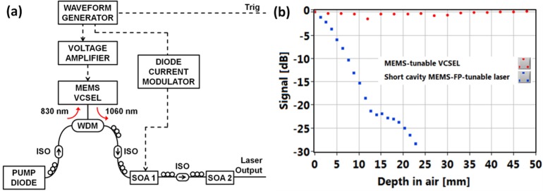
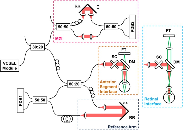
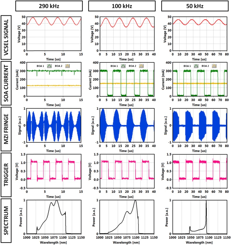
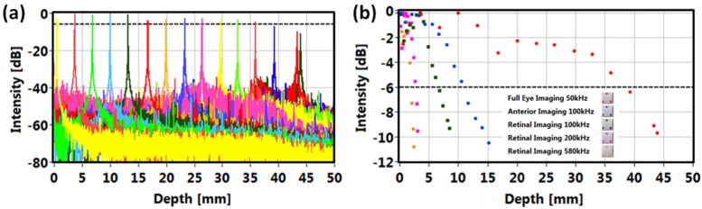
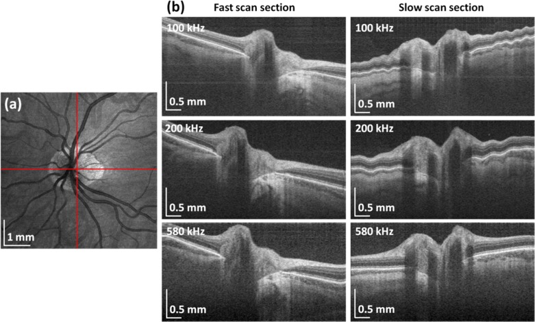
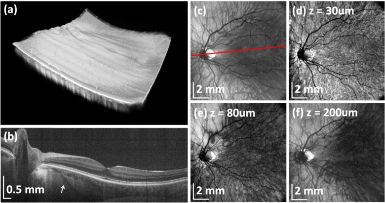
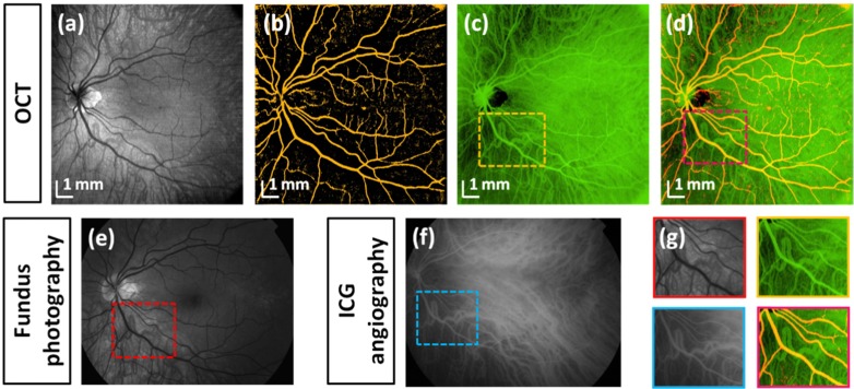
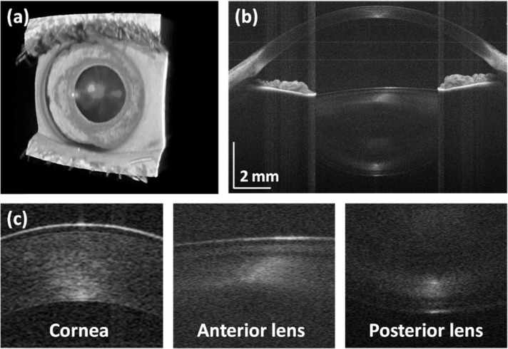
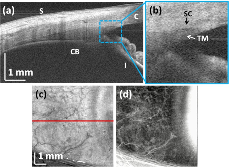
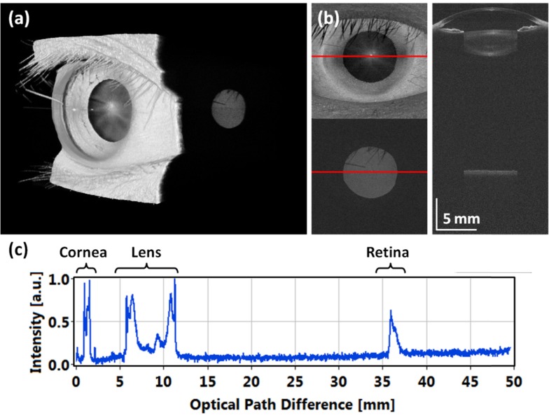
We demonstrate swept source OCT utilizing vertical-cavity surface emitting laser (VCSEL) technology for in vivo high speed retinal, anterior segment and full eye imaging. The MEMS tunable VCSEL enables long coherence length, adjustable spectral sweep range and adjustable high sweeping rate (50-580 kHz axial scan rate). These features enable integration of multiple ophthalmic applications into one instrument. The operating modes of the device include: ultrahigh speed, high resolution retinal imaging (up to 580 kHz); high speed, long depth range anterior segment imaging (100 kHz) and ultralong range full eye imaging (50 kHz). High speed imaging enables wide-field retinal scanning, while increased light penetration at 1060 nm enables visualization of choroidal vasculature. Comprehensive volumetric data sets of the anterior segment from the cornea to posterior crystalline lens surface are also shown. The adjustable VCSEL sweep range and rate make it possible to achieve an extremely long imaging depth range of ~50 mm, and to demonstrate the first in vivo 3D OCT imaging spanning the entire eye for non-contact measurement of intraocular distances including axial eye length. Swept source OCT with VCSEL technology may be attractive for next generation integrated ophthalmic OCT instruments.

Keywords: (110.4500) Optical coherence tomography; (120.4640) Optical instruments; (140.3600) Lasers, tunable; (170.4460) Ophthalmic optics and devices; (170.4470) Ophthalmology.


Figures

Fig. 1
Fig. 1
MEMS-tunable VCSEL light source and its performance. (a) Schematic of VCSEL module: WDM, wavelength-division multiplexer; ISO, isolator; SOA, booster optical amplifiers. Electrical signals indicated by dashed lines. (b) Signal vs. depth of 1060 nm swept light sources: VCSEL and short external-cavity laser.
Fig. 2
Fig. 2
Experimental setup. Retinal imaging was performed by adding ocular lens to anterior segment configuration and adjusting the fixation target path. SC – galvanometric scanners, FT – fixation target, DM – dichroic mirror, DC – dispersion compensation glass, RR – retroreflector, PDB1/PDB2 – balanced photodetectors, MZI – Mach-Zehnder interferometer.
Fig. 3
Fig. 3
Signals in VCSEL module (module driving signal, SOA current waveforms, sweep trigger), MZI fringe and integrated spectrum for selected tuning frequencies: 290 kHz, 100 kHz and 50 kHz.
Fig. 4
Fig. 4
Sensitivity roll-offs of VCSEL-OCT system: (a) point spread functions at different depths in air for the full eye imaging mode at 50 kHz, (b) signal roll-offs for different imaging modes. 6 dB signal drop is indicated by the dashed line.
Fig. 5
Fig. 5
Retinal OCT imaging using VCSEL-tunable light source. Fundus images and selected cross-sections from volumetric data sets acquired at 100 kHz (a), 200 kHz (b) and 580 kHz (c). Red-free fundus photograph indicating scanned areas at different speeds (d). Transverse sampling density and acquisition time are kept constant. Aspect ratio of all presented cross-sections is the same. High speeds enable wide-field imaging.
Fig. 6
Fig. 6
Imaging of the optic nerve head region at different speeds: fundus image (a) and extracted central fast and slow cross-sections showing reduced motion artifacts with increased speed (b).
Fig. 7
Fig. 7
Wide-field choroidal OCT imaging using VCSEL tunable light source. (a) Rendering of volumetric wide-field data set. (b) Virtual (arbitrary) cross-sectional image showing deep light penetration and ability to visualize choroid and sclera. Arrow indicates scleral vessel. (c) Projection OCT image of the entire choroid. Signal below RPE was integrated. Red line indicates direction of section in (b). OCT projection images at a depth of (d) 30 μm, (e) 80 μm and (f) 200 μm below RPE showing choroidal layers and sclera. Signal was integrated from 40 μm thick slices below RPE.
Fig. 8
Fig. 8
Wide-field OCT fundus angiography. OCT fundus image (a), segmented retinal (b) and choroidal vasculature (c), combined OCT fundus image (d). Red-free fundus photography (e). Indocyanine green (ICG) angiography (f). Comparison of details of retinal and choroidal vascular systems (g).
Fig. 9
Fig. 9
Anterior segment imaging with VCSEL-OCT: (a) rendering of the volume, (b) central averaged cross-sectional image, (c) zoomed fragments of the B-scan showing details of the corneal and the crystalline lens.
Fig. 10
Fig. 10
Corneo-scleral imaging: (a) cross-sectional OCT image presenting details of the anterior chamber angle (S – sclera, CB – ciliary body, C – cornea, I – iris); (b) zoomed portion of the image showing aqueous outflow structures (SC – Schlemm’s canal, TM – trabecular meshwork) (c) en-face view of the volumetric data set (red line indicates extracted section presented in (a)); (d) En-face visualization of scleral vessels. Scleral interface was segmented and 1 mm thick slice was integrated.
Fig. 11
Fig. 11
Full eye imaging with ultralong depth range OCT: (a) 3D rendering of volumetric data set (Media 1), (b) central cross-sectional image, (c) central B-scan extracted from data set corrected for light refraction, (d) central depth profile with echoes from the cornea, crystalline lens and the retina allows for determination of intraocular distances.

Similar articles

Cited by

References

    1. Huang D., Swanson E. A., Lin C. P., Schuman J. S., Stinson W. G., Chang W., Hee M. R., Flotte T., Gregory K., Puliafito C. A., Fujimoto J. G., “Optical coherence tomography,” Science 254(5035), 1178–1181 (1991).10.1126/science.1957169 - DOI - PMC - PubMed
    1. J. Schuman, C. A. Puliafito, and J. Fujimoto, eds., Optical Coherence Tomography of Ocular Diseases, 2nd ed. (Slack Inc., Thorofare, 2004), pp. 1–250.
    1. Wang Y., Lu A., Gil-Flamer J., Tan O., Izatt J. A., Huang D., “Measurement of total blood flow in the normal human retina using Doppler Fourier-domain optical coherence tomography,” Br. J. Ophthalmol. 93(5), 634–637 (2009).10.1136/bjo.2008.150276 - DOI - PMC - PubMed
    1. Pircher M., Hitzenberger C. K., Schmidt-Erfurth U., “Polarization sensitive optical coherence tomography in the human eye,” Prog. Retin. Eye Res. 30(6), 431–451 (2011).10.1016/j.preteyeres.2011.06.003 - DOI - PMC - PubMed
    1. Izatt J. A., Hee M. R., Swanson E. A., Lin C. P., Huang D., Schuman J. S., Puliafito C. A., Fujimoto J. G., “Micrometer-scale resolution imaging of the anterior eye in vivo with optical coherence tomography,” Arch. Ophthalmol. 112(12), 1584–1589 (1994).10.1001/archopht.1994.01090240090031 - DOI - PubMed

LinkOut - more resources