

An all-fiber-optic endoscopy platform for simultaneous OCT and fluorescence imaging
- PMID: 23162723
- PMCID: PMC3493218
- DOI: 10.1364/BOE.3.002851
An all-fiber-optic endoscopy platform for simultaneous OCT and fluorescence imaging
Abstract
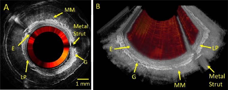
We present an all-fiber-optically based endoscope platform for simultaneous optical coherence tomography (OCT) and fluorescence imaging. This design entails the use of double-clad fiber (DCF) in the endoscope for delivery of OCT source and fluorescence excitation light while collecting the backscattered OCT signal through the single-mode core and fluorescence emission through the large inner cladding of the DCF. Circumferential beam scanning was performed by rotating a 45° reflector using a miniature DC motor at the distal end of the endoscope. Additionally, a custom DCF coupler and a wavelength division multiplexer (WDM) were utilized to seamlessly integrate both imaging modalities to achieve an entirely fiber-optically based dual-modality imaging system. We demonstrated simultaneous intraluminal 3D OCT and 2D (surface) fluorescence imaging in ex vivo rabbit esophagus using the dual-modal endomicroscopy system. Structural morphologies (provided by OCT) and fluorophore distribution (provided by the fluorescence module) could be clearly visualized, suggesting the potential of the dual-modality system for future in vivo and clinical applications.
Keywords: (170.2150) Endoscopic imaging; (170.4500) Optical coherence tomography; (170.6280) Spectroscopy, fluorescence and luminescence.
Figures


Similar articles
-
Integrated multimodal endomicroscopy platform for simultaneous en face optical coherence and two-photon fluorescence imaging.Opt Lett. 2012 Feb 1;37(3):362-4. doi: 10.1364/OL.37.000362. Opt Lett. 2012. PMID: 22297353 Free PMC article.
-
Laser tissue coagulation and concurrent optical coherence tomography through a double-clad fiber coupler.Biomed Opt Express. 2015 Mar 16;6(4):1293-303. doi: 10.1364/BOE.6.001293. eCollection 2015 Apr 1. Biomed Opt Express. 2015. PMID: 25909013 Free PMC article.
-
Dual-modality optical coherence tomography and frequency-domain fluorescence lifetime imaging microscope system for intravascular imaging.J Biomed Opt. 2020 Sep;25(9):096010. doi: 10.1117/1.JBO.25.9.096010. J Biomed Opt. 2020. PMID: 33000570 Free PMC article.
-
Double-Clad Fiber-Based Multifunctional Biosensors and Multimodal Bioimaging Systems: Technology and Applications.Biosensors (Basel). 2022 Feb 1;12(2):90. doi: 10.3390/bios12020090. Biosensors (Basel). 2022. PMID: 35200350 Free PMC article. Review.
-
Optical coherence tomography in gastroenterology: a review and future outlook.J Biomed Opt. 2017 Dec;22(12):1-17. doi: 10.1117/1.JBO.22.12.121716. J Biomed Opt. 2017. PMID: 29260538 Review.
Cited by
-
Lab-on-a-Tip (LOT): Where Nanotechnology Can Revolutionize Fibre Optics.Nanobiomedicine (Rij). 2015 Jan 1;2:3. doi: 10.5772/60518. eCollection 2015 Jan-Dec. Nanobiomedicine (Rij). 2015. PMID: 29942369 Free PMC article.
-
Circumferential optical coherence tomography angiography imaging of the swine esophagus using a micromotor balloon catheter.Biomed Opt Express. 2016 Jul 5;7(8):2927-42. doi: 10.1364/BOE.7.002927. eCollection 2016 Aug 1. Biomed Opt Express. 2016. PMID: 27570688 Free PMC article.
-
Cycloid scanning for wide field optical coherence tomography endomicroscopy and angiography in vivo.Optica. 2018 Jan 20;5(1):36-43. doi: 10.1364/OPTICA.5.000036. Optica. 2018. PMID: 29682598 Free PMC article.
-
Design and characterization of a combined OCT and wide field imaging falloposcope for ovarian cancer detection.Biomed Opt Express. 2016 Dec 8;8(1):124-136. doi: 10.1364/BOE.8.000124. eCollection 2017 Jan 1. Biomed Opt Express. 2016. PMID: 28101406 Free PMC article.
-
Focus scanning with feedback-control for fiber-optic nonlinear endomicroscopy.Biomed Opt Express. 2017 Apr 17;8(5):2519-2527. doi: 10.1364/BOE.8.002519. eCollection 2017 May 1. Biomed Opt Express. 2017. PMID: 28663888 Free PMC article.
References
-
- Li X. D., Boppart S. A., Van Dam J., Mashimo H., Mutinga M., Drexler W., Klein M., Pitris C., Krinsky M. L., Brezinski M. E., Fujimoto J. G., “Optical coherence tomography: advanced technology for the endoscopic imaging of Barrett’s esophagus,” Endoscopy 32(12), 921–930 (2000).10.1055/s-2000-9626 - DOI - PubMed
-
- Jang I. K., Bouma B. E., Kang D. H., Park S. J., Park S. W., Seung K. B., Choi K. B., Shishkov M., Schlendorf K., Pomerantsev E., Houser S. L., Aretz H. T., Tearney G. J., “Visualization of coronary atherosclerotic plaques in patients using optical coherence tomography: comparison with intravascular ultrasound,” J. Am. Coll. Cardiol. 39(4), 604–609 (2002).10.1016/S0735-1097(01)01799-5 - DOI - PubMed
-
- Hanna N., Saltzman D., Mukai D., Chen Z., Sasse S., Milliken J., Guo S., Jung W., Colt H., Brenner M., “Two-dimensional and 3-dimensional optical coherence tomographic imaging of the airway, lung, and pleura,” J. Thorac. Cardiovasc. Surg. 129(3), 615–622 (2005).10.1016/j.jtcvs.2004.10.022 - DOI - PubMed
Grants and funding
LinkOut - more resources
Full Text Sources
Other Literature Sources