. 2012 Sep 1;1(6):829-839.
doi: 10.4161/onci.20492.

High endothelial venules (HEVs) in human melanoma lesions: Major gateways for tumor-infiltrating lymphocytes

Affiliations

High endothelial venules (HEVs) in human melanoma lesions: Major gateways for tumor-infiltrating lymphocytes

Ludovic Martinet et al. Oncoimmunology. .

Abstract

The presence of tumor-infiltrating lymphocytes (TILs) is a strong prognostic parameter for local dissemination and overall survival in melanoma. Lymphocyte migration from blood into peripheral tissues is mainly regulated by vascular endothelium. However, the blood vessels and mechanisms governing the recruitment of TILs in melanoma tumors remain poorly understood. Here, we show that high endothelial venules (HEVs), specialized blood vessels for lymphocyte extravasation into lymphoid tissues, are frequently found in melanoma tumors and are associated with high levels of lymphocyte infiltration. The analysis of 225 primary melanomas revealed that lymphocytes specifically infiltrated HEV-rich areas of melanoma tumors and that the density of MECA-79+ HEVs was variable among patients and strongly correlated with CD3+, CD8+ and CD20+ TIL densities. Inflammatory (CCL5, CXCL9, CXCL10 and CXCL11) and lymphoid (CCL21, CCL19 and CXCL13) chemokines as well as TH1 and naïve T-cell genes were overexpressed in melanoma samples with high densities of tumor HEVs. Mature dendritic cells (mDCs) were frequently found around tumor HEVs and densities of HEVs and DC-LAMP+ mDCs within tumor stroma were strongly correlated. DCs which maintain HEVs in lymph nodes, may thus also contribute to the regulation of HEVs in melanomas. Finally, we found significantly higher densities of tumor HEVs in melanomas with tumor regression, low Clark level of invasion and thin Breslow thickness (all p < 0.001). The strong association between tumor HEVs, TILs, mDCs and clinical parameters of melanoma, supports a critical role for HEVs in limiting malignant melanoma development through both naïve and effector T-lymphocyte recruitment and activation.


Figures
None
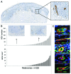
Figure 1. Human melanomas contain variable numbers of HEV blood vessels. (A) Representative picture from a digitized tumor slide stained with MECA-79 antibody showing numerous HEV blood vessels located at the invasive front of a melanoma lesion (dashed area). (B) Histograms showing the absolute number and the density of MECA-79+ HEVs in the tumor area quantified for 225 primary melanomas. Two representative pictures of melanomas with high and low numbers of MECA-79+ HEVs are shown. (C) Phenotypic characterization of tumor HEVs. Tumor HEVs express pan-endothelial cell markers CD31 and vWB, post-capillary venule-specific marker DARC and HEV-specific markers MECA-79, HECA-452, G72 and G152. Immunofluorescence staining of melanoma tumor sections was performed with the indicated antibodies. Counterstaining was performed with DAPI.
None
Figure 2. The density of tumor HEVs correlates with clinical characteristics and prognostic biomarkers of primary melanoma. The clinical characteristics and prognostic biomarkers of 225 primary melanomas were analyzed according to the density of HEVs. Significant differences in the density of tumor HEVs were found between patients classified according to melanoma histologic types (A), tumor regression (B), Breslow thickness (C) and Clark level of invasion (D). The line in the center of each box represents the median value of the distribution, and the upper and lower ends of the box are the upper and lower quartiles, respectively. Comparisons between groups were performed using the Kruskal-wallis test.
None
Figure 3. Phenotypic characterization of immune populations associated with HEV blood vessels in melanoma tumors. (A) Immune infiltrates are located around MECA-79+ HEV (red)-rich areas of melanoma lesions (dashed area). (B) MECA-79+ HEVs (green) are surrounded by numerous CD3+ T lymphocytes (red) that infiltrate melan-A+ tumors (blue). (C) CD3+ T cells (red) are seen attached to the luminal surface of MECA-79+ HEV endothelial cells (green) and extravasating through the vessel wall. (D) HEVs are present in tumor areas infiltrated mainly by CD3+ T cells (green) and CD20+ B cells (red). (E) A considerable fraction of CD3+ T cells (green) surrounding MECA-79+ HEVs (blue) are CD8+ T cytotoxic T cells (red).
None
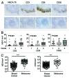
Figure 4. The density of tumor HEVs predicts lymphocyte infiltration in melanoma. (A, B) Consecutive tumor sections from 225 melanomas were analyzed by immunohistochemistry with MECA-79, anti-CD3, anti-CD8 and anti-CD20 antibodies. The number of CD3+, CD8+ and CD20+ TILs was quantified by semi quantitative optical grading (grade 1, 2, 3 for low, moderate and high density of positive cells). (A) Representative pictures showing HEVs and the indicated immune populations in tumors with (upper panel) and without (lower panel) tumor HEVs. (B) Graphs showing the density of HEVs in melanoma tumors according to the density of CD3+, CD8+ and CD20+ TILs. p < 0.001; Kruskal-wallis test. (C) Graphs showing the densities of HEVs and CD3+ T cells (lower graph) in melanomas (n = 225) and invasive breast tumors (n = 146). *** p < 0.001; Mann Whitney test.
None
Figure 5. The density of tumor HEVs has no influence on the density of tumor infiltrating Foxp3+ T cells. (A) Representative immunofluorescence staining of melanoma tumor sections with the indicated antibodies showing Foxp3+ T cells (red) within T cells infiltrates (green) in melanomas containing MECA-79+ HEVs (blue) or not. (B) Consecutive melanoma sections from representative tumors with a high and a low density of MECA-79+ HEVs (n = 30) were stained with anti-Foxp3 antibodies and the density of Foxp3+ cells (red arrow) within melanoma lymphoid stroma was calculated. (C) The density of Foxp3+ cells within peri-tumor lymphoid infiltrates is not correlated with the density of tumor HEVs. (D) The density of Foxp3+ cells within peri-tumor lymphoid infiltrates is similar in melanomas with a low and a high density of tumor HEVs. n.s p > 0.05 Mann Whitney test.
None
Figure 6. Expression of specific chemokines, chemokine receptors, TH1 and naïve T cell genes in melanoma tumors containing HEVs. (A) Expression of genes related to lymphocyte migration was determined by qRT-PCR in 14 melanoma samples according to the presence of tumor HEVs (7 HEV+ vs 7 HEV-). Relative mRNA expression levels were adjusted to the levels of the housekeeping gene YWHAZ, and are represented as mean relative expression (+SD) for HEV+ (red bars) and HEV- (blue bars) tumors. (B) Representative immunofluorescence staining of melanoma tumor sections with the indicated antibodies showing numerous CD45RO+ lymphocytes and a few CD45RA+ naïve T cells around MECA-79+ HEVs. (C) Expression of genes related to T Helper orientation was determined by qRT-PCR in 14 melanoma samples according to the presence of tumor HEVs (7 HEV+ vs 7 HEV-). The levels are represented as mean relative expression (+SD) for HEV+ (red bars) and HEV- (blue bars) tumors. * p < 0.05, ** p < 0.01, *** p < 0.001; Mann Whitney test.
None
Figure 7. The presence of tumor HEVs in melanoma lesions is correlated with the number of DC-LAMP+ DCs clusters. (A) Immunofluorescence staining showing Fascin+ DCs (red) around MECA-79+ HEVs (green) within melanoma lymphoid infiltrates. (B-D) Consecutive melanoma sections from tumors with a high and a low density of MECA-79+ HEVs (n = 30) were stained with antibodies directed against DC-LAMP and the number of DC-LAMP+ cells clusters was calculated. (B) Representative picture showing MECA-79+ HEVs in a tumor area infiltrated by DC-LAMP+ DCs. (C) The density of MECA-79+ HEVs is significantly higher in melanomas with a high amount of DC-LAMP+ infiltrating DCs (grades 2–3) as compared with melanomas with a low amount of DC-LAMP+ infiltrating DCs (grade 1). (D) The number of DC-LAMP+ DCs clusters is significantly higher in melanomas with a high density of tumor HEVs. *** p < 0.001; Mann Whitney test.

References

    1. MacKie RM, Hauschild A, Eggermont AM. Epidemiology of invasive cutaneous melanoma. Ann Oncol. 2009;20(Suppl 6):vi1–7. doi: 10.1093/annonc/mdp252. - DOI - PMC - PubMed
    1. Disis ML. Immune regulation of cancer. J Clin Oncol. 2010;28:4531–8. doi: 10.1200/JCO.2009.27.2146. - DOI - PMC - PubMed
    1. Pagès F, Berger A, Camus M, Sanchez-Cabo F, Costes A, Molidor R, et al. Effector memory T cells, early metastasis, and survival in colorectal cancer. N Engl J Med. 2005;353:2654–66. doi: 10.1056/NEJMoa051424. - DOI - PubMed
    1. Galon J, Costes A, Sanchez-Cabo F, Kirilovsky A, Mlecnik B, Lagorce-Pagès C, et al. Type, density, and location of immune cells within human colorectal tumors predict clinical outcome. Science. 2006;313:1960–4. doi: 10.1126/science.1129139. - DOI - PubMed
    1. Zhang L, Conejo-Garcia JR, Katsaros D, Gimotty PA, Massobrio M, Regnani G, et al. Intratumoral T cells, recurrence, and survival in epithelial ovarian cancer. N Engl J Med. 2003;348:203–13. doi: 10.1056/NEJMoa020177. - DOI - PubMed

Publication types