

Tumor cells, rather than dendritic cells, deliver antigen to the lymph node for cross-presentation
- PMID: 23162751
- PMCID: PMC3489739
- DOI: 10.4161/onci.20493
Tumor cells, rather than dendritic cells, deliver antigen to the lymph node for cross-presentation
Abstract
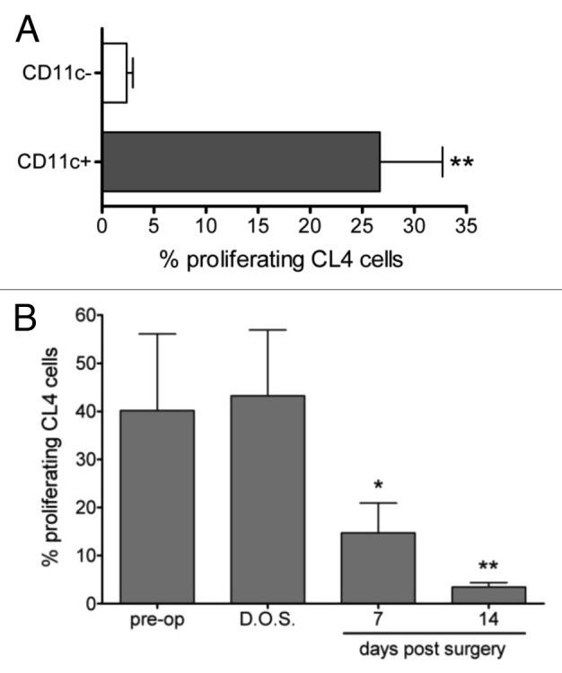
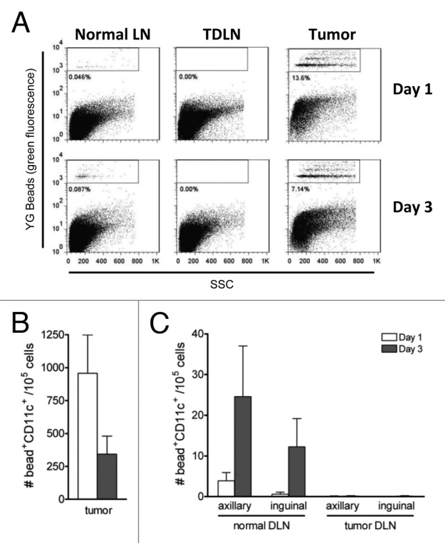
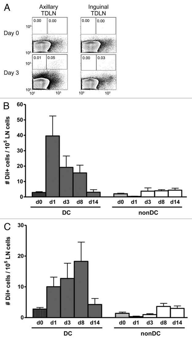
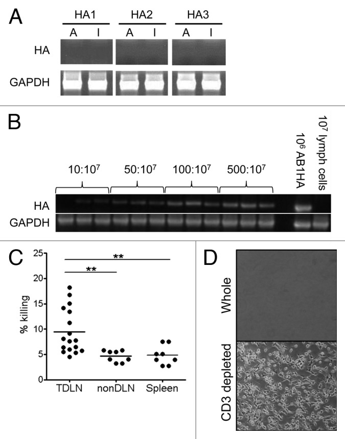
It is widely accepted that generation of tumor specific CD8+ T-cell responses occur via cross-priming; however the source of tumor antigen for this event is unknown. We examined the source and form of tumor antigen required for cross-presentation in the local lymph node (LN) using a syngeneic mouse tumor model expressing a marker antigen. We found that cross-presentation of this model tumor antigen in the LN is dependent on continuous traffic of antigen from the tumor site, but without any detectable migration of tumor resident dendritic cells (DCs). Instead, small numbers of tumor cells metastasize to local LNs where they are exposed to a localized CTL attack, resulting in delivery of tumor antigen into the cross-presentation pathway.
Figures
Similar articles
-
Migratory dendritic cells transfer antigen to a lymph node-resident dendritic cell population for efficient CTL priming.Immunity. 2006 Jul;25(1):153-62. doi: 10.1016/j.immuni.2006.04.017. Immunity. 2006. PMID: 16860764
-
Spreading the load: Antigen transfer between migratory and lymph node-resident dendritic cells promotes T-cell priming.Eur J Immunol. 2017 Oct;47(10):1798-1801. doi: 10.1002/eji.201747248. Eur J Immunol. 2017. PMID: 28845904
-
Delivered antigen peptides to resident CD8α+ DCs in lymph node by micelle-based vaccine augment antigen-specific CD8+ effector T cell response.Eur J Pharm Biopharm. 2020 Feb;147:76-86. doi: 10.1016/j.ejpb.2019.12.013. Epub 2019 Dec 28. Eur J Pharm Biopharm. 2020. PMID: 31887349
-
Dendritic cell gene therapy.Surg Oncol Clin N Am. 2002 Jul;11(3):645-60. doi: 10.1016/s1055-3207(02)00027-3. Surg Oncol Clin N Am. 2002. PMID: 12487060 Review.
-
Unique functions of splenic CD8alpha+ dendritic cells during infection with intracellular pathogens.Immunol Lett. 2007 Dec 15;114(2):66-72. doi: 10.1016/j.imlet.2007.09.007. Epub 2007 Oct 12. Immunol Lett. 2007. PMID: 17964665 Review.
Cited by
-
The role and therapeutic implications of T cells in cancer of the lung.Clin Transl Immunology. 2019 Aug 28;8(8):e1076. doi: 10.1002/cti2.1076. eCollection 2019. Clin Transl Immunology. 2019. PMID: 31485330 Free PMC article.
-
Predicted limited redistribution of T cells to secondary lymphoid tissue correlates with increased risk of haematological malignancies in asplenic patients.Sci Rep. 2021 Aug 12;11(1):16394. doi: 10.1038/s41598-021-95225-x. Sci Rep. 2021. PMID: 34385480 Free PMC article.
-
Immunosuppressive Mechanisms of Malignant Gliomas: Parallels at Non-CNS Sites.Front Oncol. 2015 Jul 6;5:153. doi: 10.3389/fonc.2015.00153. eCollection 2015. Front Oncol. 2015. PMID: 26217588 Free PMC article. Review.
-
Leveraging Endogenous Dendritic Cells to Enhance the Therapeutic Efficacy of Adoptive T-Cell Therapy and Checkpoint Blockade.Front Immunol. 2020 Sep 25;11:578349. doi: 10.3389/fimmu.2020.578349. eCollection 2020. Front Immunol. 2020. PMID: 33101304 Free PMC article. Review.
-
Sentinel Lymph Node in Non-Small Cell Lung Cancer: Assessment of Feasibility and Safety by Near-Infrared Fluorescence Imaging and Clinical Consequences.J Pers Med. 2022 Dec 30;13(1):90. doi: 10.3390/jpm13010090. J Pers Med. 2022. PMID: 36675751 Free PMC article.
References
-
- Marzo AL, Lake RA, Lo D, Sherman L, McWilliam A, Nelson D, et al. Tumor antigens are constitutively presented in the draining lymph nodes. J Immunol. 1999;162:5838–45. - PubMed
-
- van Mierlo GJD, Boonman ZFHM, Dumortier HMH, den Boer AT, Fransen MF, Nouta J, et al. Activation of dendritic cells that cross-present tumor-derived antigen licenses CD8+ CTL to cause tumor eradication. J Immunol. 2004;173:6753–9. - PubMed
LinkOut - more resources
Full Text Sources
Other Literature Sources
Research Materials