Sustaining attention to simple tasks: a meta-analytic review of the neural mechanisms of vigilant attention
- PMID: 23163491
- PMCID: PMC3627747
- DOI: 10.1037/a0030694
Sustaining attention to simple tasks: a meta-analytic review of the neural mechanisms of vigilant attention
Abstract
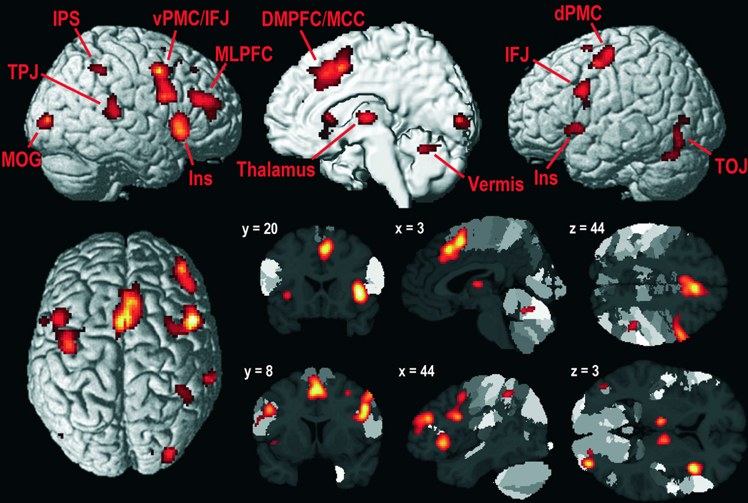
Maintaining attention for more than a few seconds is essential for mastering everyday life. Yet, our ability to stay focused on a particular task is limited, resulting in well-known performance decrements with increasing time on task. Intriguingly, such decrements are even more likely if the task is cognitively simple and repetitive. The attentional function that enables our prolonged engagement in intellectually unchallenging, uninteresting activities has been termed vigilant attention. Here we synthesized what we have learned from functional neuroimaging about the mechanisms of this essential mental faculty. To this end, a quantitative meta-analysis of pertinent neuroimaging studies was performed, including supplementary analyses of moderating factors. Furthermore, we reviewed the available evidence on neural time-on-task effects, additionally considering information obtained from patients with focal brain damage. Integrating the results of both meta-analysis and review, we identified a set of mainly right-lateralized brain regions that may form the core network subserving vigilant attention in humans, including dorsomedial, mid- and ventrolateral prefrontal cortex, anterior insula, parietal areas (intraparietal sulcus, temporoparietal junction), and subcortical structures (cerebellar vermis, thalamus, putamen, midbrain). We discuss the potential functional roles of different nodes of this network as well as implications of our findings for a theoretical account of vigilant attention. It is conjectured that sustaining attention is a multicomponent, nonunitary mental faculty, involving a mixture of (a) sustained/recurrent processes subserving task-set/arousal maintenance and (b) transient processes subserving the target-driven reorienting of attention. Finally, limitations of previous studies are considered and suggestions for future research are provided.
2013 APA, all rights reserved
Figures


References
-
- Adler CM, Sax KW, Holland SK, Schmithorst V, Rosenberg L, Strakowski SM. Changes in neuronal activation with increasing attention demand in healthy volunteers: an fMRI study. Synapse. 2001;42:266–272. - PubMed
-
- Alegria J. The time course of preparation after a first peak: Some constraints of reacting mechanisms. Quarterly Journal of Experimental Psychology. 1974;26:622–632.
-
- Alexander MP, Stuss DT, Shallice T, Picton TW, Gillingham S. Impaired concentration due to frontal lobe damage from two distinct lesion sites. Neurology. 2005;65:572–579. - PubMed
-
- Allen M. Models of hemispheric specialization. Psychological Bulletin. 1983;93:73–104. - PubMed
-
- Amunts K, Malikovic A, Mohlberg H, Schormann T, Zilles K. Brodmann's areas 17 and 18 brought into stereotaxic space-where and how variable? NeuroImage. 2000;11:66–84. - PubMed
Publication types
MeSH terms
Grants and funding
LinkOut - more resources
Full Text Sources
Other Literature Sources