

High expression of KIBRA in low atypical protein kinase C-expressing gastric cancer correlates with lymphatic invasion and poor prognosis
- PMID: 23163744
- PMCID: PMC7657249
- DOI: 10.1111/cas.12066
High expression of KIBRA in low atypical protein kinase C-expressing gastric cancer correlates with lymphatic invasion and poor prognosis
Abstract
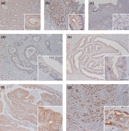
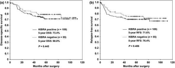
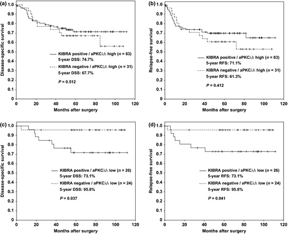
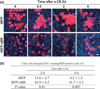
Overexpression of atypical protein kinase Cλ/ι (aPKCλ/ι), a regulator of cell polarity, is frequently associated with the poor prognoses of several cancers, including gastric cancer. Recent studies revealed a molecular link between aPKC and KIBRA, an upstream regulator of tumor suppressor Hippo pathway that regulates cell proliferation and apoptosis. Further, KIBRA directly inhibits the kinase activity of aPKC to regulate epithelial cell polarity. These observations suggest that the KIBRA-aPKC connection plays a role in cancer progression; however, clinical significance of the correlation between these factors remains unclear. Here we examined the correlation between KIBRA/aPKCλ/ι expression, as detected by immunohistochemistry, and clinicopathological outcomes in 164 gastric cancer patients using Fisher's exact test and Kaplan-Meier log-rank test. We found an intimate correlation between the expression level of KIBRA and aPKCλ/ι (P = 0.012). Furthermore, high expression of KIBRA is correlated with lymphatic (P = 0.046) and venous invasion (P = 0.039). The expression level of KIBRA by itself did not correlate with the prognosis; however, high expression of KIBRA in low aPKCλ/ι-expressing gastric cancer correlated with disease-specific (P = 0.037) and relapse-free survival (P = 0.041) by Kaplan-Meier with log-rank test and higher lymphatic invasion cases by Fisher's exact test (P = 0.042). Furthermore, overexpression of the aPKC-binding region of KIBRA disrupted tight junctions in epithelial cells. These results suggest that high expression of KIBRA in low aPKC-expressing cells causes massive loss of aPKC activity, leading to loss of polarity and invasiveness of gastric cancer cells.
© 2012 Japanese Cancer Association.
Figures
Similar articles
-
KIBRA suppresses apical exocytosis through inhibition of aPKC kinase activity in epithelial cells.Curr Biol. 2011 Apr 26;21(8):705-11. doi: 10.1016/j.cub.2011.03.029. Epub 2011 Apr 14. Curr Biol. 2011. PMID: 21497093
-
High expression of atypical protein kinase C lambda/iota in gastric cancer as a prognostic factor for recurrence.Ann Surg Oncol. 2010 Jan;17(1):81-8. doi: 10.1245/s10434-009-0708-x. Epub 2009 Sep 23. Ann Surg Oncol. 2010. PMID: 19774416
-
Elevated expression of Par3 promotes prostate cancer metastasis by forming a Par3/aPKC/KIBRA complex and inactivating the hippo pathway.J Exp Clin Cancer Res. 2017 Oct 10;36(1):139. doi: 10.1186/s13046-017-0609-y. J Exp Clin Cancer Res. 2017. PMID: 29017577 Free PMC article.
-
The Par3/Par6/aPKC complex and epithelial cell polarity.Exp Cell Res. 2013 Jun 10;319(10):1357-64. doi: 10.1016/j.yexcr.2013.03.021. Epub 2013 Mar 25. Exp Cell Res. 2013. PMID: 23535009 Review.
-
Protein kinase Cλ/ι in cancer: a contextual balance of time and signals.Trends Cell Biol. 2022 Dec;32(12):1023-1034. doi: 10.1016/j.tcb.2022.04.002. Epub 2022 Apr 29. Trends Cell Biol. 2022. PMID: 35501226 Free PMC article. Review.
Cited by
-
WWC Proteins: Important Regulators of Hippo Signaling in Cancer.Cancers (Basel). 2021 Jan 15;13(2):306. doi: 10.3390/cancers13020306. Cancers (Basel). 2021. PMID: 33467643 Free PMC article. Review.
-
Apical polarity and actomyosin dynamics control Kibra subcellular localization and function in Drosophila Hippo signaling.Dev Cell. 2023 Oct 9;58(19):1864-1879.e4. doi: 10.1016/j.devcel.2023.08.029. Epub 2023 Sep 19. Dev Cell. 2023. PMID: 37729921 Free PMC article.
-
Low expression of WWC1, a tumor suppressor gene, is associated with aggressive breast cancer and poor survival outcome.FEBS Open Bio. 2019 Jul;9(7):1270-1280. doi: 10.1002/2211-5463.12659. Epub 2019 Jun 1. FEBS Open Bio. 2019. PMID: 31102318 Free PMC article.
-
KIBRA attains oncogenic activity by repressing RASSF1A.Br J Cancer. 2017 Jun 29;117(4):553-62. doi: 10.1038/bjc.2017.192. Online ahead of print. Br J Cancer. 2017. PMID: 28664913 Free PMC article.
-
KIBRA promotes prostate cancer cell proliferation and motility.FEBS J. 2016 May;283(10):1800-11. doi: 10.1111/febs.13718. Epub 2016 Apr 5. FEBS J. 2016. PMID: 27220053 Free PMC article.
References
-
- Jemal A, Siegel R, Ward E et al Cancer statistics, 2006. CA Cancer J Clin 2006; 56: 106–30. - PubMed
-
- Henson DE, Dittus C, Younes M, Nguyen H, Albores‐Saavedra J. Differential trends in the intestinal and diffuse types of gastric carcinoma in the United States, 1973–2000: increase in the signet ring cell type. Arch Pathol Lab Med 2004; 128: 765–70. - PubMed
-
- Faycal J, Bessaguet C, Nousbaum JB et al Epidemiology and long term survival of gastric carcinoma in the French district of Finistere between 1984 and 1995. Gastroenterol Clin Biol 2005; 29: 23–32. - PubMed
-
- Cunningham SC, Kamangar F, Kim MP et al Survival after gastric adenocarcinoma resection: eighteen‐year experience at a single institution. J Gastrointest Surg 2005; 9: 718–25. - PubMed
-
- Tsubono Y, Hisamichi S. Screening for gastric cancer in Japan. Gastric Cancer 2000; 3: 9–18. - PubMed
Publication types
MeSH terms
Substances
LinkOut - more resources
Full Text Sources
Other Literature Sources
Medical
