. 2012 Dec;86(6):1424-40.
doi: 10.1111/mmi.12066. Epub 2012 Nov 19.

The response threshold of Salmonella PilZ domain proteins is determined by their binding affinities for c-di-GMP

Affiliations

The response threshold of Salmonella PilZ domain proteins is determined by their binding affinities for c-di-GMP

Ingrid Swanson Pultz et al. Mol Microbiol. 2012 Dec.

Abstract

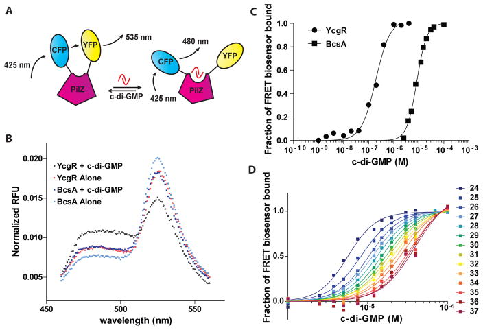
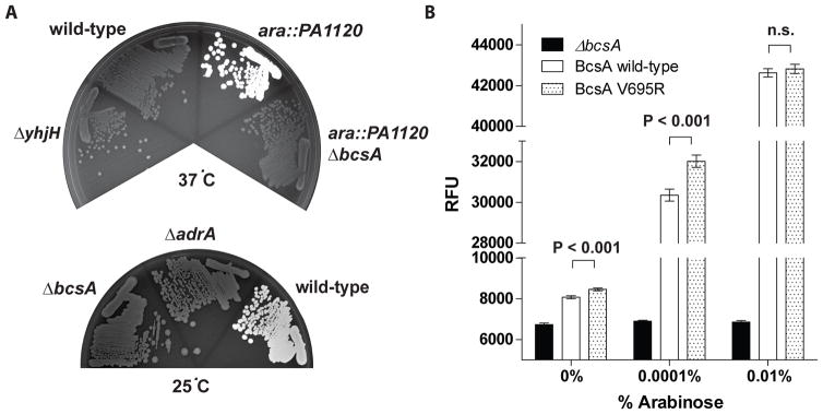
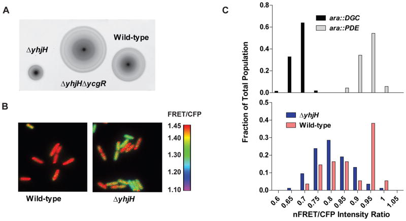
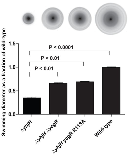
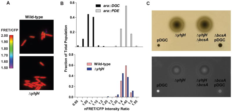
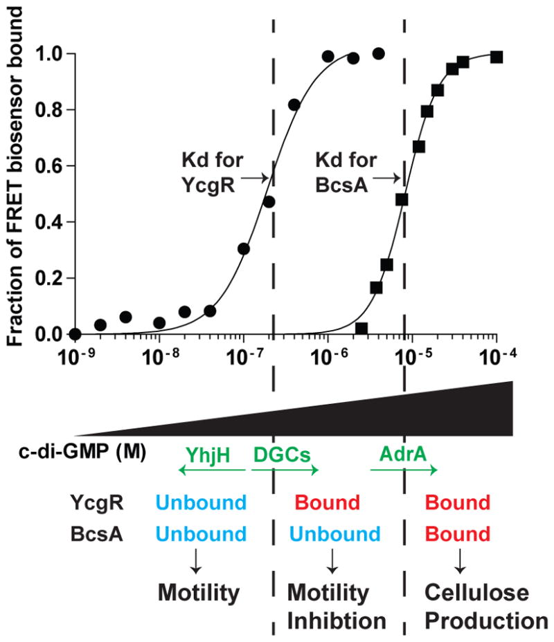
c-di-GMP is a bacterial second messenger that is enzymatically synthesized and degraded in response to environmental signals. Cellular processes are affected when c-di-GMP binds to receptors which include proteins that contain the PilZ domain. Although each c-di-GMP synthesis or degradation enzyme metabolizes the same molecule, many of these enzymes can be linked to specific downstream processes. Here we present evidence that c-di-GMP signalling specificity is achieved through differences in affinities of receptor macromolecules. We show that the PilZ domain proteins of Salmonella Typhimurium, YcgR and BcsA, demonstrate a 43-fold difference in their affinity for c-di-GMP. Modulation of the affinities of these proteins altered their activities in a predictable manner in vivo. Inactivation of yhjH, which encodes a predicted c-di-GMP degrading enzyme, increased the fraction of the cellular population that demonstrated c-di-GMP levels high enough to bind to the higher-affinity YcgR protein and inhibit motility, but not high enough to bind to the lower-affinity BcsA protein and stimulate cellulose production. Finally, PilZ domain proteins of Pseudomonas aeruginosa demonstrated a 145-fold difference in binding affinities, suggesting that regulation by binding affinity may be a conserved mechanism that allows organisms with many c-di-GMP binding macromolecules to rapidly integrate multiple environmental signals into one output.


Figures

Fig. 1
Fig. 1. The binding properties of the BcsA FRET construct for c-di-GMP
A. PilZ domain FRET proteins were generated by the fusion of the fluorophores mYPet (YFP) and mCyPet (CFP) to the termini of a PilZ domain protein. These FRET fusion proteins demonstrate a characteristic FRET signal when exposed to the excitation wavelength of mCyPet (425 nm). Binding of c-di-GMP to the PilZ domain changes the conformation of the PilZ domain protein, which results in a shift in the emission ratio of mYPet (535 nm) to mCyPet (480 nm). In this figure, binding of c-di-GMP results in a decrease in FRET, though c-di-GMP binding can also result in a FRET increase depending on the PilZ domain protein used. B. Normalized fluorescence emission spectra of YcgR and BcsA based FRET biosensors upon excitation of mCyPet at 425 nm in vitro. Shown are the emission spectra of the YcgR biosensor in the presence (black circles) and absence (red circles) of 40 μM c-di-GMP, and the BcsA biosensor in the presence (dark blue squares) and absence (light blue squares) of 40 μM c-di-GMPat 25°C. For both biosensors, the addition of c-di- GMP results in a decrease in the amount of FRET. C. c-di-GMP binding curves for YcgR (circles) and BcsA (squares) biosensors at 25°C as measured by FRET. The fraction of FRET biosensor bound to c-di-GMP was calculated as follows: Fraction of FRET biosensor bound = (FRET ratio(x) - FRET ratio free) / (FRET ratio bound - FRET ratio free). A number of 0 indicates that the FRET biosensor is completely unbound to c-di-GMP, and a number of 1 indicates that the FRET biosensor is completely bound to c-di-GMP. D. Binding curves of the BcsA biosensor at temperatures ranging from 24°C to 37°C at 1°C intervals as indicated by the legend. Binding affinity decreases as temperature increases.
Fig. 2
Fig. 2. Mutation of the residue at Position-X alters the c-di-GMP binding affinities of S. Typhimurium PilZ domain proteins
A. Table showing the c-di-GMP switch region for YcgR and BcsA. Shown in white are mutated residues. Small arrows show the locations of the invariant arginines of the RxxxR motif. The affinities shown are for proteins measured at 25°C. B. c-di-GMP binding curves for YcgR wild-type (open circles) and YcgR R113A (closed circles) FRET biosensors at 25°C. C. c-di-GMP binding curves for BcsA PilZ wild-type (open squares) and BcsA PilZ V695R (closed squares) FRET biosensors at 25°C.
Fig. 3
Fig. 3. c-di-GMP binding affinity of S. Typhimurium BcsA affects cellulose production in vivo
A. S. Typhimurium makes cellulose at 24°C, but not at 37°C, in a manner that is dependent on both AdrA and BcsA. Calcofluor agar plates incubated at either 37°C (top) or 24°C (bottom) with wild-type, ΔyhjH, ara::DGC, ara::DGC ΔbcsA, ΔbcsA, or ΔadrA strains of S. Typhimurium. ara::DGC is a strain that harbors an arabinose-inducible DGC on the chromosome. The plate incubated at 37°C was supplemented with 0.001% arabinose. Plates were exposed to UV light with an excitation wavelength of 365 nm, which is a wavelength that stimulates fluorescence of calcofluor when it is bound to cellulose. White coloring indicates calcofluor binding to cellulose. B. Calcofluor fluorescence at different concentrations of arabinose at 37°C for ara::DGC with wild-type bcsA, or ara::PA1120 in which the bcsA gene has either been deleted (ΔbcsA) or mutated (BcsA V695R). Shown is the average fluorescence emission intensity at 420 nm, after excitation at 365 nm, of cells growing on 200 μl agar in individual wells of a 96-well plate in a representative experiment. RFU, relative fluorescence units. n.s. = not significant.
Fig. 4
Fig. 4. c-di-GMP binding affinity of S. Typhimurium YcgR affects motility inhibition
A. Swimming halos on motility agar plates with 25 μM IPTG of S. Typhimurium strains harboring pDGC or pDGC-YcgR at 37°C for 12 hours. A wild-type strain without exogenous DGC expression was not shown since the swimming diameter of this strain is too large to include and adequately show the differences between strains harboring pDGC. B. Swimming diameters of ΔycgR harboring either pDGC-YcgR or pDGC-YcgR R113A relative to the swimming diameter of the uncomplemented ΔycgR pDGC strain, at indicated levels of IPTG at 37°C. Both YcgR and YcgR R113A effect a decrease in motility at increasing IPTG levels, but the YcgR R113A mutant protein requires a higher concentration of IPTG to inhibit motility than the wild-type YcgR protein.
Fig. 5
Fig. 5. A S. Typhimurium strain harboring a yhjH mutation demonstrates a larger proportion of cells with c-di-GMP bound to YcgR than the wild-type strain
A. Swimming halos on motility agar plates of wild-type, ΔyhjH, and ΔyhjHΔycgR at 37°C. B. Dual-emission ratio microscopic images (FRET/CFP) of S. Typhimurium (above) or a ΔyhjH mutant (below) expressing pYcgR-Spy. Pseudocolors represent emission ratios (527/480 nm) of the FRET-based biosensor as indicated by the figure legend to the right. C. Histogram showing the fraction of cells that demonstrate the indicated average nFRET/CFP ratios for either control strains expressing a DGC or a PDE off of an arabinose-inducible promoter (top), or for wild-type or yhjH mutant strains (bottom). nFRET/CFP ratios differ from FRET/CFP ratios since nFRET intensities have been corrected for bleedthrough and fluorescence in the YFP channel. The difference between the wild-type and ΔyhjH strains is highly significant (P<0.0001). nFRET: net FRET intensity, calculated by subtracting bleedthrough coefficients and intensity of the YFP channel as detailed in Materials and Methods. Over 100 cells were analyzed for each strain.
Fig. 6
Fig. 6. A S. Typhimurium strain harboring the lower-affinity ycgR-R113A mutation does not demonstrate YcgR-dependent motility inhibition in a ΔyhjH mutant
Above: swimming diameters (from left to right) of ΔyhjH, ΔyhjHΔycgR, ΔyhjH ycgR-R113A, and wild-type S. Typhimurium, on motility agar plates incubated at 37°C for 9 hours. Below: quantitation of the swimming diameters of indicated S. Typhimurium strains compared to the wild-type strain. Statistical significance is indicated.
Fig. 7
Fig. 7. A S. Typhimurium ΔyhjH strain does not demonstrate c-di-GMP binding to the BcsA PilZ domain or cellulose production
A. Dual-emission ratio microscopic images (FRET/CFP) of wild-type (top) or ΔyhjH mutant (bottom) S. Typhimurium expressing pBcsA-Spy. Pseudocolors represent emission ratios (527/480 nm) of the FRET-based biosensor as indicated by the figure legend to the left. B. Histogram showing the fraction of cells that demonstrate the indicated nFRET/CFP ratios for control strains expressing a DGC or a PDE off of an arabinose-inducible promoter (top) and either wild-type or ΔyhjH mutant strains (bottom). nFRET/CFP ratios are different than FRET/CFP ratios since nFRET intensities have been corrected for bleedthrough and fluorescence in the YFP channel. nFRET: net FRET intensity, calculated by subtracting bleedthrough coefficients and intensity of the YFP channel as detailed in Materials and Methods. The difference in nFRET/CFP ratios between the wild-type and the ΔyhjH is not significant (P > 0.05). C. Visible light (top) and UV (bottom) images of strains of S. Typhimurium on soft- agar motility plates with calcofluor, incubated at 37°C for 2 hours. This plate was supplemented with 100 mM IPTG.
Fig. 8
Fig. 8. The binding affinities of YcgR and BcsA for c-di-GMP determines cellular phenotypes
When cellular c-di-GMP levels are kept low by PDEs such as YhjH, neither PilZ domain protein is bound to c-di- GMP, resulting in a motile cell that does not produce cellulose. As cellular DGCs increase the concentration of c-di-GMP past the Kd for YcgR, YcgR becomes bound to c-di-GMP and thus inhibits motility, even at the levels of c-di-GMP that are not high enough to bind BcsA. Activation of AdrA expression results in enough c-di-GMP being produced to bind to BcsA, and cellulose synthesis occurs.

References

    1. Amikam D, Galperin MY. PilZ domain is part of the bacterial c-di-GMP binding protein. Bioinformatics. 2006;22:3–6. - PubMed
    1. Benach J, Swaminathan SS, Tamayo R, Handelman SK, Folta-Stogniew E, Ramos JE, Forouhar F, Neely H, Seetharaman J, Camilli A, Hunt JF. The structural basis of cyclic diguanylate signal transduction by PilZ domains. EMBO J. 2007;26:5153–5166. - PMC - PubMed
    1. Bochner BR, Huang HC, Schieven GL, Ames BN. Positive selection for loss of tetracycline resistance. J Bacteriol. 1980;143:926–933. - PMC - PubMed
    1. Boehm A, Kaiser M, Li H, Spangler C, Kasper CA, Ackermann M, Kaever V, Sourjik V, Roth V, Jenal U. Second messenger-mediated adjustment of bacterial swimming velocity. Cell. 2010;141:107–116. - PubMed
    1. Christen B, Christen M, Paul R, Schmid F, Folcher M, Jenoe P, Meuwly M, Jenal U. Allosteric control of cyclic di-GMP signaling. J Biol Chem. 2006;281:32015–32024. - PubMed

Publication types

MeSH terms