. 2012 Nov 19:12:266.
doi: 10.1186/1471-2180-12-266.

Adaptive response to starvation in the fish pathogen Flavobacterium columnare: cell viability and ultrastructural changes

Affiliations

Adaptive response to starvation in the fish pathogen Flavobacterium columnare: cell viability and ultrastructural changes

Covadonga R Arias et al. BMC Microbiol. .

Abstract

Background: The ecology of columnaris disease, caused by Flavobacterium columnare, is poorly understood despite the economic losses that this disease inflicts on aquaculture farms worldwide. Currently, the natural reservoir for this pathogen is unknown but limited data have shown its ability to survive in water for extended periods of time. The objective of this study was to describe the ultrastructural changes that F. columnare cells undergo under starvation conditions. Four genetically distinct strains of this pathogen were monitored for 14 days in media without nutrients. Culturability and cell viability was assessed throughout the study. In addition, cell morphology and ultrastructure was analyzed using light microscopy, scanning electron microscopy, and transmission electron microscopy. Revival of starved cells under different nutrient conditions and the virulence potential of the starved cells were also investigated.

Results: Starvation induced unique and consistent morphological changes in all strains studied. Cells maintained their length and did not transition into a shortened, coccus shape as observed in many other Gram negative bacteria. Flavobacterium columnare cells modified their shape by morphing into coiled forms that comprised more than 80% of all the cells after 2 weeks of starvation. Coiled cells remained culturable as determined by using a dilution to extinction strategy. Statistically significant differences in cell viability were found between strains although all were able to survive in absence of nutrients for at least 14 days. In later stages of starvation, an extracellular matrix was observed covering the coiled cells. A difference in growth curves between fresh and starved cultures was evident when cultures were 3-months old but not when cultures were starved for only 1 month. Revival of starved cultures under different nutrients revealed that cells return back to their original elongated rod shape upon encountering nutrients. Challenge experiments shown that starved cells were avirulent for a fish host model.

Conclusions: Specific morphological and ultrastructural changes allowed F. columnare cells to remain viable under adverse conditions. Those changes were reversed by the addition of nutrients. This bacterium can survive in water without nutrients for extended periods of time although long-term starvation appears to decrease cell fitness and resulted in loss of virulence.


Figures
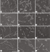
Figure 1
Figure 1
Morphology of Flavobacterium columnare cells during starvation in ultrapure water as determined by SEM. Panels A, B, and C display ATCC 23643 strain. Panels D, E, and F show ARS-1 strain. Panels G, H, I show ALG-00-530 strain. Panels J, K, and L display ALG-02-36 strain. Panels A, D, G, and J show cells at day 1 (scale bar 10 μm); panels B, E, H, and K display 7 days starved cells (scale bar 5 μm); panels C, F, I, and L show 14 day starved cells (scale bar 1 μm).
Figure 2
Figure 2
Percent of bacillus and coiled forms observed over time during starvation in ultrapure water. Bacillus and coiled forms are represented by solid and open symbols, respectively. ARS-1 (■), ALG-00-530 (●), and ALG-02-36 (▲).
Figure 3
Figure 3
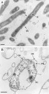
TEM observations of Flavobacterium columnare ALG-00-530 strain in ultrapure water. Panel A, day 1 after transfer to ultrapure water. Panel B, maintained in ultrapure water for 150 days. Arrows indicate surface blebbing (SB), membrane vesicle (MV), nucleoid (N), cell membrane (CM), outer membrane (OM), periplasmic space (PS), inclusion (I), and nucleoid compaction areas (NC). Scale bars represent 500 nm.
Figure 4
Figure 4
Flavobacterium columnare ALG-00-530 strain after starvation in ultrapure water for 150 days as determined by SEM. Arrow indicates the only bacillus observed in this preparation. Scale bar represents 1 μm.
Figure 5
Figure 5
Growth curves of 24-h (♦), 1-month (□), and 3-month () old cultures of strain ALG-00-530 cultivated in MS at 28°C. Data points represent means and error bars represent standard errors.
Figure 6
Figure 6
Growth curves of 5-month old Flavobacterium columnare ALG-00-530 cultures incubated under different nutrient conditions. Modified Sheih (MS) medium (■), diluted MS (MS-10) (□), MS without yeast extract (MS-T) (○), MS without tryptone (MS-Y) (), and MS without nutrients (MS-S) (▼). Data points represent means and error bars represent standard errors.
Figure 7
Figure 7
Morphology changes of Flavobacterium columnare starved cells during revival in different nutrient media. Panels A and B, cells cultured in Modified Sheih (MS) medium at 4 h post-inoculation (arrows point to small membrane vesicles). Panel C, a cell cultured in diluted MS (MS-10) at 4 h post-inoculation (arrow indicates fimbriae). Panel D, active cells division observed in MS-10 cultures at 12 h post-inoculation. Panel E, cells actively growing in MS at 36 h post-inoculation displaying membrane vesicles (arrow). Panel F, coiled forms (arrow) observed in MS-10 cultures at 36 h post-inoculation. Scale bars represent 1 μm.

Similar articles

Cited by

References

    1. Austin B, Austin DA. Bacterial fish pathogens: disease of farmed and wild fish. New York, NY: Springer; 1999.
    1. Wagner BA, Wise DJ, Khoo LH, Terhune JS. The epidemiology of bacterial diseases in food-size channel catfish. J Aquat Anim Heal. 2002;14:263–272. doi: 10.1577/1548-8667(2002)014<0263:TEOBDI>2.0.CO;2. - DOI - PubMed
    1. Figueiredo HCP, Klesius PH, Arias CR, Evans J, Shoemaker CA, Pereira DJ, Peixoto MTD. Isolation and characterization of strains of Flavobacterium columnare from Brazil. J Fish Dis. 2005;28(4):199–204. doi: 10.1111/j.1365-2761.2005.00616.x. - DOI - PubMed
    1. Amin NE, Abdallah IS, Faisal M, Easa ME, Alaway T, Alyan SA. Columnaris infection among cultured Nile tilapia Oreochromis niloticus. Antonie Van Leeuwenhoek J Microbiol. 1988;54(6):509–520. doi: 10.1007/BF00588387. - DOI - PubMed
    1. Decostere A, Haesebrouck F, Van Driessche E, Charlier G, Ducatelle R. Characterization of the adhesion of Flavobacterium columnare (Flexibacter columnaris) to gill tissue. J Fish Dis. 1999;22:465–474. doi: 10.1046/j.1365-2761.1999.00198.x. - DOI

Publication types

LinkOut - more resources