

Phosphatidylserine exposure on the surface of Leishmania amazonensis amastigotes modulates in vivo infection and dendritic cell function
- PMID: 23163958
- PMCID: PMC3565004
- DOI: 10.1111/pim.12019
Phosphatidylserine exposure on the surface of Leishmania amazonensis amastigotes modulates in vivo infection and dendritic cell function
Abstract
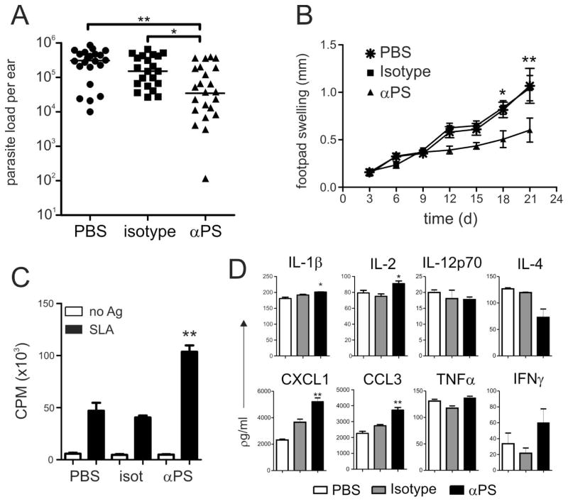
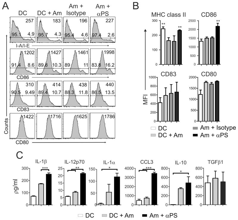
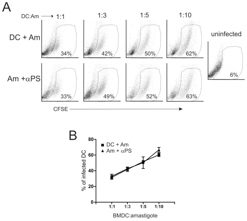
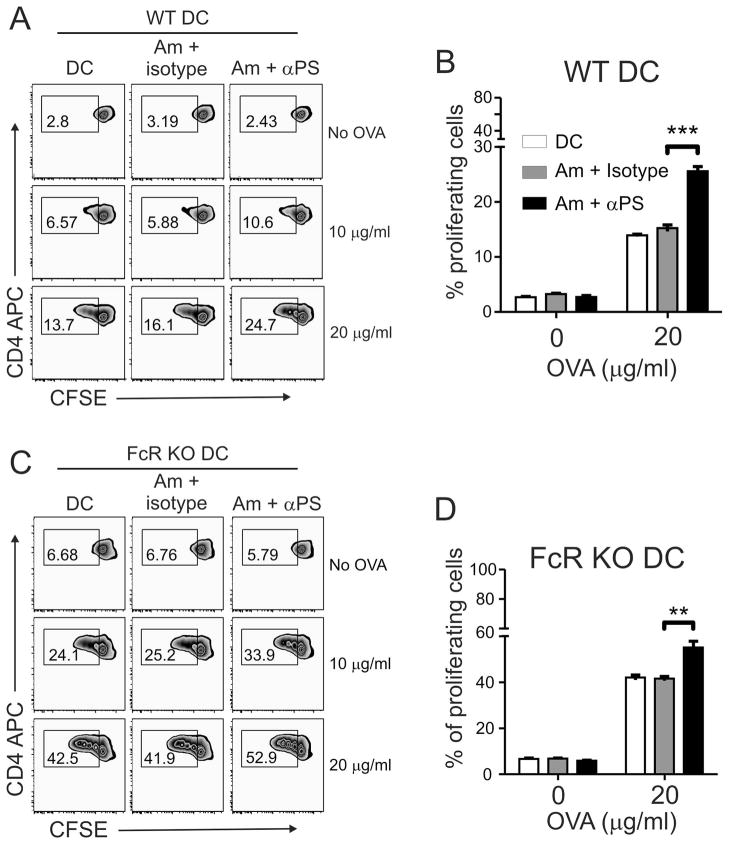
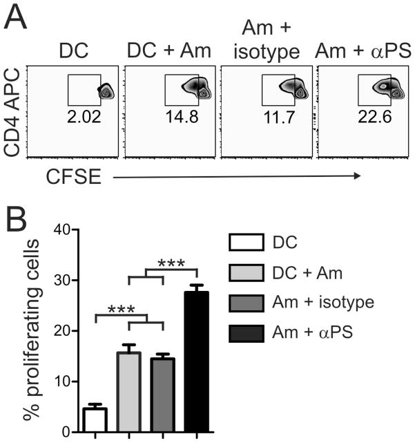
Leishmania amazonensis parasites can cause diverse forms of leishmaniasis in humans and persistent lesions in most inbred strains of mice. In both cases, the infection is characterized by a marked immunosuppression of the host. We previously showed that amastigote forms of the parasite make use of surface-exposed phosphatidylserine (PS) molecules to infect host cells and promote alternative macrophage activation, leading to uncontrolled intracellular proliferation of the parasites. In this study, we demonstrated that treatment of infected mice with a PS-targeting monoclonal antibody ameliorated parasite loads and lesion development, which correlated with increased proliferative responses by lymphocytes. In addition, we observed an enhanced dendritic cell (DC) activation and antigen presentation in vitro. Our data imply that the recognition of PS exposed on the surface of amastigotes plays a role in down-modulating DC functions, in a matter similar to that of apoptotic cell clearance. This study provides new information regarding the mechanism of immune suppression in Leishmania infection.
© 2012 Blackwell Publishing Ltd.
Figures
Similar articles
-
CD4+ T Cell-Dependent Macrophage Activation Modulates Sustained PS Exposure on Intracellular Amastigotes of Leishmania amazonensis.Front Cell Infect Microbiol. 2019 Apr 12;9:105. doi: 10.3389/fcimb.2019.00105. eCollection 2019. Front Cell Infect Microbiol. 2019. PMID: 31032234 Free PMC article.
-
Leishmania amazonensis Subverts the Transcription Factor Landscape in Dendritic Cells to Avoid Inflammasome Activation and Stall Maturation.Front Immunol. 2020 Jun 9;11:1098. doi: 10.3389/fimmu.2020.01098. eCollection 2020. Front Immunol. 2020. PMID: 32582184 Free PMC article.
-
Mimicry of apoptotic cells by exposing phosphatidylserine participates in the establishment of amastigotes of Leishmania (L) amazonensis in mammalian hosts.J Immunol. 2006 Feb 1;176(3):1834-9. doi: 10.4049/jimmunol.176.3.1834. J Immunol. 2006. PMID: 16424214
-
Modulation of dendritic cell function by Leishmania parasites.J Immunol. 2008 Apr 1;180(7):4355-60. doi: 10.4049/jimmunol.180.7.4355. J Immunol. 2008. PMID: 18354154 Free PMC article. Review.
-
Interaction of Leishmania parasites with dendritic cells and its functional consequences.Immunobiology. 2004;209(1-2):173-7. doi: 10.1016/j.imbio.2004.02.007. Immunobiology. 2004. PMID: 15481151 Review.
Cited by
-
Apoptotic mimicry as a strategy for the establishment of parasitic infections: parasite- and host-derived phosphatidylserine as key molecule.Cell Commun Signal. 2020 Jan 15;18(1):10. doi: 10.1186/s12964-019-0482-8. Cell Commun Signal. 2020. PMID: 31941500 Free PMC article. Review.
-
The Apoptotic Role of Metacaspase in Toxoplasma gondii.Front Microbiol. 2016 Jan 19;6:1560. doi: 10.3389/fmicb.2015.01560. eCollection 2015. Front Microbiol. 2016. PMID: 26834715 Free PMC article.
-
Bacterial imaging and photodynamic inactivation using zinc(II)-dipicolylamine BODIPY conjugates.Photochem Photobiol Sci. 2015 Jul;14(7):1271-81. doi: 10.1039/c5pp00100e. Photochem Photobiol Sci. 2015. PMID: 26063101 Free PMC article.
-
CD300a Receptor Blocking Enhances Early Clearance of Leishmania donovani From Its Mammalian Host Through Modulation of Effector Functions of Phagocytic and Antigen Experienced T Cells.Front Immunol. 2022 Jan 18;12:793611. doi: 10.3389/fimmu.2021.793611. eCollection 2021. Front Immunol. 2022. PMID: 35116028 Free PMC article.
-
Bidirectional cytokine-microRNA control: A novel immunoregulatory framework in leishmaniasis.PLoS Pathog. 2022 Aug 4;18(8):e1010696. doi: 10.1371/journal.ppat.1010696. eCollection 2022 Aug. PLoS Pathog. 2022. PMID: 35925884 Free PMC article. Review.
References
-
- Reithinger R, Dujardin JC, Louzir H, Pirmez C, Alexander B, Brooker S. Cutaneous leishmaniasis. Lancet Infect Dis. 2007;7:581–596. - PubMed
-
- Jones DE, Buxbaum LU, Scott P. IL-4-independent inhibition of IL-12 responsiveness during Leishmania amazonensis infection. J Immunol. 2000;165:364–372. - PubMed
-
- Buxbaum LU, Uzonna JE, Goldschmidt MH, Scott P. Control of New World cutaneous leishmaniasis is IL-12 independent but STAT4 dependent. Eur J Immunol. 2002;32:3206–3215. - PubMed
Publication types
MeSH terms
Substances
Grants and funding
LinkOut - more resources
Full Text Sources
Other Literature Sources
