Review
. 2012 Nov 20:7:56.
doi: 10.1186/1750-1326-7-56.

Regulated protein aggregation: stress granules and neurodegeneration

Affiliations
Review

Regulated protein aggregation: stress granules and neurodegeneration

Benjamin Wolozin. Mol Neurodegener. .

Abstract

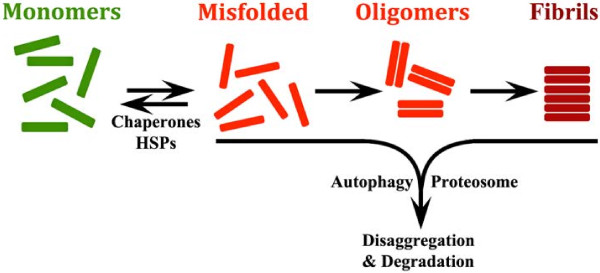
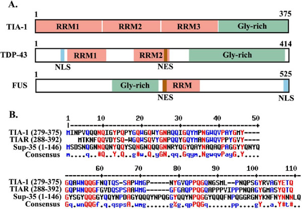
The protein aggregation that occurs in neurodegenerative diseases is classically thought to occur as an undesirable, nonfunctional byproduct of protein misfolding. This model contrasts with the biology of RNA binding proteins, many of which are linked to neurodegenerative diseases. RNA binding proteins use protein aggregation as part of a normal regulated, physiological mechanism controlling protein synthesis. The process of regulated protein aggregation is most evident in formation of stress granules. Stress granules assemble when RNA binding proteins aggregate through their glycine rich domains. Stress granules function to sequester, silence and/or degrade RNA transcripts as part of a mechanism that adapts patterns of local RNA translation to facilitate the stress response. Aggregation of RNA binding proteins is reversible and is tightly regulated through pathways, such as phosphorylation of elongation initiation factor 2α. Microtubule associated protein tau also appears to regulate stress granule formation. Conversely, stress granule formation stimulates pathological changes associated with tau. In this review, I propose that the aggregation of many pathological, intracellular proteins, including TDP-43, FUS or tau, proceeds through the stress granule pathway. Mutations in genes coding for stress granule associated proteins or prolonged physiological stress, lead to enhanced stress granule formation, which accelerates the pathophysiology of protein aggregation in neurodegenerative diseases. Over-active stress granule formation could act to sequester functional RNA binding proteins and/or interfere with mRNA transport and translation, each of which might potentiate neurodegeneration. The reversibility of the stress granule pathway also offers novel opportunities to stimulate endogenous biochemical pathways to disaggregate these pathological stress granules, and perhaps delay the progression of disease.


Figures

Figure 1
Figure 1
The conventional model for degenerative disease based on mass action and hydrophobic interactions. Monomeric proteins randomly misfold. The chaperone system, including heat shock proteins (HSPs), can reverse the misfolding, and produce normal, functional proteins. However, the misfolded proteins are prone to random oligomerization, and evidence suggests that the resulting oligomers can be toxic. The oligomers aggregate further to form fibrillar aggregates. In each case formation of the misfolded proteins, oligomers and fibrils are considered to lack normal biological functions. These oligomers and fibrillar aggregates can be removed by degradation, which occurs through the actions of the autophagic system and the ubiquitin proteasomal system. Increasing evidence suggests that autophagy is the predominant mechanism of degradation in diseases such as Alzheimer’s disease and Parkinson’s disease [3].
Figure 2
Figure 2
Structures of RNA binding proteins.A) RNA binding proteins, such as TIA-1, TDP-43 and FUS, contain RNA-recognition motifs (RRM), which bind RNA and Glycine rich (Gly-rich) domains that mediate protein aggregation. TDP-43 and FUS contain discrete nuclear localization and nuclear export signals (NLS, NES), while nuclear localization of TIA-1 does not appear to localize to particular domains [11,12]. B) Alignment of glycine rich domains of TIA-1, TIAR and Sup-35. The alignment was performed using the “multalin” program (http://multalin.toulouse.inra.fr). In the consensus sequence, the red letters correspond to homologous amino acids, where the # symbol is used to convey imperfect homologies, and the blue letters refer to partial homology.
Figure 3
Figure 3
Structure and functions of RNA binding proteins. RNA binding proteins have dual sites of action. In the nucleus, many RNA binding proteins, such as TDP-43, SMN (SMN1 and 2), TIA-1 and FUS regulate mRNA splicing. RNA binding proteins are also present in the cytoplasm and neuronal arbors, where they regulate RNA transport, activity dependent protein synthesis and sequestration of unnecessary transcripts in response to stress. Each of the RNA binding proteins shown in the figure associate with stress granules. TIA-1, SMN and Pumillio-2 are important for trafficking of mRNA in axons and dendrites, which is mediated by microtubules (blue and mustard striped line) and molecular motors. At the synapse, different RNA binding proteins regulate activity dependent translation. Phosphorylation causes 4E-BP to dissociate from eIF4E, which initiates translation. FMRP inhibits this process; loss of FMRP expression (such as occurs in fragile X syndrome) leads to excessive synaptic protein synthesis and excessive dendritic spine density. In contrast, CPEB stimulates activity dependent translation in a process that might involve regulated protein aggregation [27]. Activity dependent protein synthesis is modulated by microRNA. For instance, miR125 regulates the synthesis of mGluR and PSD-95 [24]. miRNA are generated by action of the RISC complex and argonaute, which cleave precursors to generate the miRs. Adapted from Liu-Yesucevitz, et al. [10].
Figure 4
Figure 4
Mechanism of normal and pathological stress granule formation.A) In normal, physiological conditions, neurons synthesize specialized proteins from capped transcripts. The proteins eIF4A, E and G complex to form the eIF4F pre-initiation complex, which interacts with the ribosome (40S) as well as other translational regulators to synthesize proteins. Association with the 60S ribosome complex allows protein synthesis to begin. B) Stress leads to phosphorylation of eIF2α, dissociation of ribosomes and many of the translation initiation factors, leaving mRNA bound eIF4G and poly-A binding protein. Nucleating RNA binding proteins bind the free RNA and also form protein/protein complexes, which initiate stress granule formation. Once initiated, other RNA binding proteins bind to the mRNA and to the nucleating RNA binding proteins to increase the size and complexity of SGs. These SGs are rapidly reversible upon removal of the stress, however prolonged SG formation affects cell biology by interacting with biological systems regulating apoptosis, signaling and RNA decay. C) Pathological proteins, such as TDP-43, FUS and tau, have a strong tendency to form oligomers, and then fibrils. The consolidation of RNA binding proteins during SG formation might promote oligomerization by creating cellular domains with higher concentrations of these proteins. Conversely, the increased stability of oligomers and fibrils might serve as a nidus for SG formation, leading to over-active SG formation. Microtubule associated protein tau also participates in this process because it mislocates to the soma and dendritic arbor leading to interactions with SG proteins and potentially stimulating SG formation [41]. Tau also directly binds RNA [42].

References

    1. Dobson CM. Protein folding and misfolding. Nature. 2003;426:884–890. doi: 10.1038/nature02261. - DOI - PubMed
    1. Jarrett J, Jarrett J, Lansbury P. Seeding "one-dimensional crystallization" of amyloid: a pathogenic mechanism in Alzheimer's disease and scrapie? Cell. 1993;73(P Lansbury):1055–1058. - PubMed
    1. Bandhyopadhyay U, Cuervo AM. Chaperone-mediated autophagy in aging and neurodegeneration: lessons from alpha-synuclein. Exp Gerontol. 2006;42:120–128. - PubMed
    1. Powers ET, Morimoto RI, Dillin A, Kelly JW, Balch WE. Biological and chemical approaches to diseases of proteostasis deficiency. Annu Rev Biochem. 2009;78:959–991. doi: 10.1146/annurev.biochem.052308.114844. - DOI - PubMed
    1. Selkoe DJ, Schenk D. Alzheimer's disease: molecular understanding predicts amyloid-based therapeutics. Annu Rev Pharmacol Toxicol. 2003;43:545–584. doi: 10.1146/annurev.pharmtox.43.100901.140248. - DOI - PubMed

Publication types

MeSH terms