. 2012 Nov 19:12:265.
doi: 10.1186/1471-2180-12-265.

Role of the small GTPase Rab27a during herpes simplex virus infection of oligodendrocytic cells

Affiliations

Role of the small GTPase Rab27a during herpes simplex virus infection of oligodendrocytic cells

Raquel Bello-Morales et al. BMC Microbiol. .

Abstract

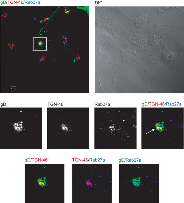
Background: The morphogenesis of herpes simplex virus type 1 (HSV-1) comprises several events, of which some are not completely understood. It has been shown that HSV-1 glycoproteins accumulate in the trans-Golgi network (TGN) and in TGN-derived vesicles. It is also accepted that HSV-1 acquires its final morphology through a secondary envelopment by budding into TGN-derived vesicles coated with viral glycoproteins and tegument proteins. Nevertheless, several aspects of this process remain elusive. The small GTPase Rab27a has been implicated in regulated exocytosis, and it seems to play a key role in certain membrane trafficking events. Rab27a also seems to be required for human cytomegalovirus assembly. However, despite the involvement of various Rab GTPases in HSV-1 envelopment, there is, to date, no data reported on the role of Rab27a in HSV-1 infection.

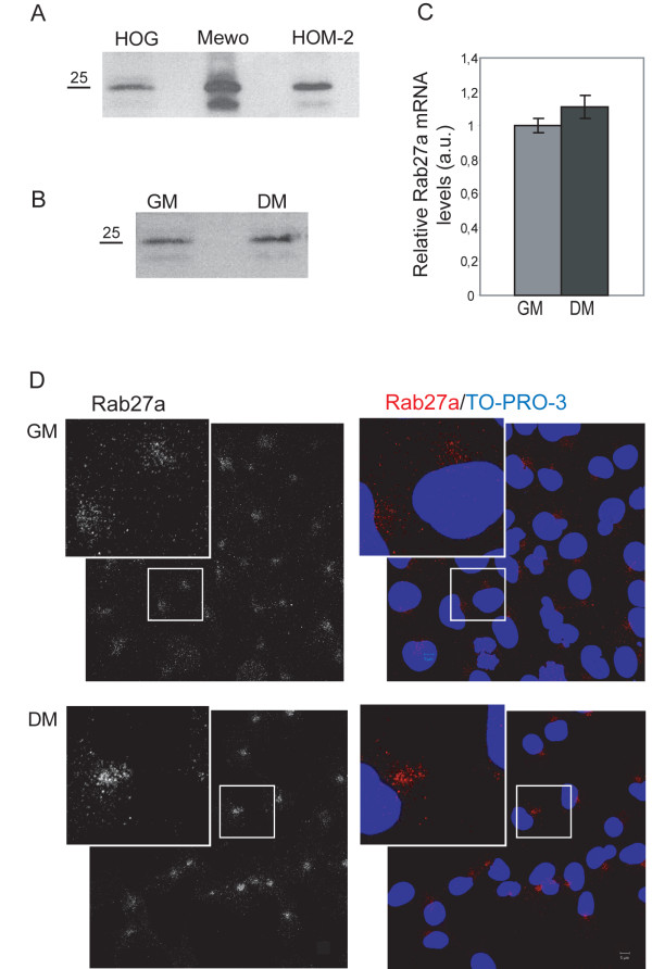
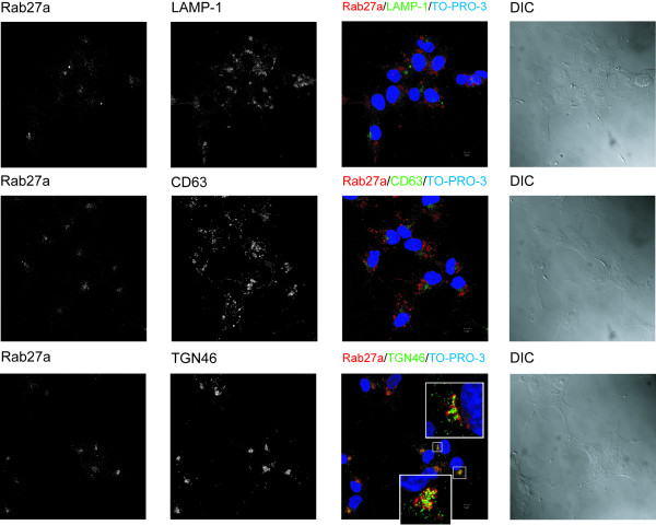
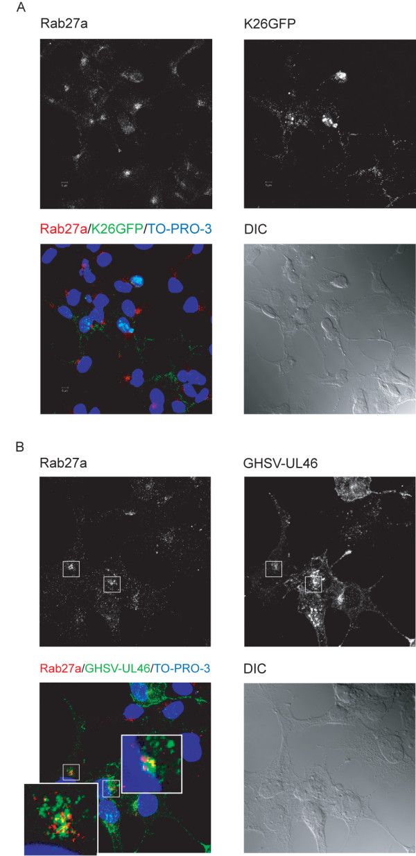
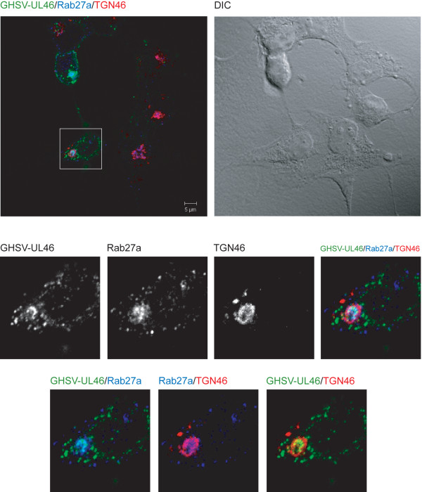
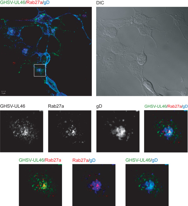
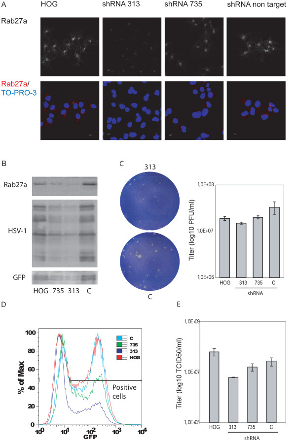
Results: Herein, we show that Rab27a colocalized with GHSV-UL46, a tegument-tagged green fluorescent protein-HSV-1, in the TGN. In fact, this small GTPase colocalized with viral glycoproteins gH and gD in that compartment. Functional analysis through Rab27a depletion showed a significant decrease in the number of infected cells and viral production in Rab27a-silenced cells.

Conclusions: Altogether, our results indicate that Rab27a plays an important role in HSV-1 infection of oligodendrocytic cells.


Figures

Figure 1
Figure 1
Expression of Rab27a in HOG cell line.A. HOG cells cultured in GM were subjected to SDS–PAGE under non-reducing conditions and analyzed by immunoblotting with anti-Rab27a polyclonal antibody. Compared to positive controls, Mewo and HOM-2 cell lines, HOG cells show significant levels of Rab27a expression. B. RTqPCR quantification of relative Rab27a mRNA expression levels in HOG cells cultured in GM or DM. C. Immunoblot analysis of Rab27a expression in HOG cells cultured in GM or DM. HOG cells were subjected to SDS–PAGE under non-reducing conditions and analyzed by immunoblotting with anti-Rab27a polyclonal antibody Immunoblot assays showed a moderate increase of Rab27a in DM cultures. D. HOG cells cultured in GM or DM were fixed and processed for confocal immunofluorescence analysis with anti-Rab27a polyclonal antibody, detected using an Alexa Fluor 555 secondary antibody. The squares correspond to enlarged regions showing pericentrosomal localization of Rab27a, more scattered in the case of GM cultures. Images correspond to the projection of the planes obtained by confocal microscopy. (DIC: Differential Interference Contrast). All data are representative of, at least, 3 independent experiments. (a.u., arbitrary units).
Figure 2
Figure 2
Subcellular localization of Rab27a in HOG cells. A. HOG cells cultured in DM were fixed and processed for confocal double-label indirect immunofluorescence analysis with anti-Rab27a polyclonal antibody and antibodies against LAMP-1, CD63 and TGN-46. Primary antibodies were detected using Alexa Fluor 555 and 488 secondary antibodies. Images correspond to the projection of the planes obtained by confocal microscopy. Colocalization (yellow spots) was detected between Rab27a and TGN-46. The squares show enlarged images corresponding to a confocal slice of 0.8 μm. (DIC: Differential Interference Contrast).
Figure 3
Figure 3
Expression and localization of Rab27a in HSV-1-infected cells. Triple-label indirect confocal immunofluorescence analysis of HOG cells infected with K26GFP (A) or GHSV-UL46 (B) with an anti-Rab27a polyclonal antibody, detected with an Alexa Fluor 555 secondary antibody. The squares show enlarged images corresponding to a confocal slice of 0.8 μm showing partial colocalization of GHSV-UL46 with Rab27a (yellow spots). (DIC: Differential Interference Contrast).
Figure 4
Figure 4
Colocalization between GHSV-UL46 and Rab27a in the TGN. HOG cells cultured in DM and infected at a m.o.i. of 1 with GHSV-UL46 were fixed and processed for confocal triple-label indirect immunofluorescence analysis with anti-Rab27a and anti-TGN-46 polyclonal antibodies. Low panels, corresponding to confocal slices of 0.8 μm, are enlargements of the square shown in upper panel, which corresponds to the projection of the planes obtained by confocal microscopy. Images show colocalization between Rab27a and GHSV-UL46 in the TGN. Colocalization between Rab27a and GHSV-UL46 appears cyan; between Rab27a and TGN, magenta; between GHSV-UL46 and the TGN, yellow; colocalization between Rab27a, GHSV-UL46 and TGN appears white. (DIC: Differential Interference Contrast).
Figure 5
Figure 5
Colocalization between Rab27a and gD. HOG cells cultured in DM and infected at a m.o.i. of 1 with GHSV-UL46 were fixed and processed for confocal triple-label indirect immunofluorescence analysis with polyclonal anti-Rab27a and anti-gD LP2 antibodies. Low panels, corresponding to confocal slices of 0.8 μm, are enlargements of the squares shown in upper panels, which correspond to the projection of the planes obtained by confocal microscopy. Images show colocalization between Rab27a and gD. Colocalization between Rab27a and GHSV-UL46 appears yellow; between Rab27a and gD, magenta; between GHSV-UL46 and gD, cyan; colocalization between Rab27a, GHSV-UL46 and gD appears white. (DIC: Differential Interference Contrast).
Figure 6
Figure 6
Colocalization between Rab27a and viral glycoproteins in the TGN. HOG cells cultured in DM and infected at a m.o.i. of 1 with wild-type HSV-1 were fixed and processed for confocal triple-label indirect immunofluorescence analysis with polyclonal anti-Rab27a and anti-TGN-46 antibodies. Low panels, corresponding to confocal slices of 0.8 μm, are enlargements of the squares shown in upper panels, which correspond to the projection of the planes obtained by confocal microscopy. Colocalization between gD and the TGN appears yellow; between Rab27a and the TGN, magenta; between Rab27a and gD, cyan. Arrow points to colocalization of Rab27a with gD in the TGN. (DIC: Differential Interference Contrast).
Figure 7
Figure 7
Effect of Rab27a depletion on HSV-1 infection. HOG cells mock-transfected or transfected with Rab27a-silencing shRNA-313 or shRNA-735, and shRNA non target control, were fixed and processed for confocal immunofluorescence analysis with polyclonal anti-Rab27a antibody. As images show, shRNA-313 induced an efficient knockdown of Rab27a while shRNA-735 produced a weaker effect (A). Equal number of cells were subjected to SDS–PAGE and analyzed by immunoblotting with anti-HSV-1 and anti-GFP antibodies. In Rab27a-depleted cells, a significant decrease in viral-associated GFP signal can be observed (B). Plaque assay shows a drastic reduction in plaque size and a decrease in the viral production determined by the number of plaque forming units (p.f.u.) per ml in silenced shRNA-313 cells compared to control cells (C). Silenced cells and controls infected at a m.o.i. of 1 with K26GFP were processed for flow cytometry, analyzing fluorescence of GFP (D). Percentage (%) of max designates the number of cells relative to the maximum fraction. For each fluorescence intensity within positive cells, the percentage of silenced cells corresponding to shRNA-313 and 735 is considerably lower than controls. Data are representative of 3 independent experiments. E. Rab27a-depleted cells and controls were infected at a m.o.i. of 0.5 with HSV-1. 18 h p.i., viral titers were determined by TCID50. Virus yield was significantly reduced in shRNA-313 silenced cells.

References

    1. Noseworthy JH. Progress in determining the causes and treatment of multiple sclerosis. Nature. 1999;399:A40–A47. - PubMed
    1. Christensen T. Human herpesviruses in MS. Int MS j/MS Forum. 2007;14:41–47. - PubMed
    1. Sanders VJ, Waddell AE, Felisan SL, Li X, Conrad AJ, Tourtellotte WW. Herpes simplex virus in postmortem multiple sclerosis brain tissue. Arch Neurol. 1996;53:125–133. doi: 10.1001/archneur.1996.00550020029012. - DOI - PubMed
    1. Charpin C, Gambarelli D, Lavaut MN, Seigneurin JM, Raphael M, Berard M, Toga M. Herpes simplex virus antigen detection in human acute encephalitis: an immunohistochemical study using avidin-biotin-peroxidase complex method. Acta neuropathol. 1985;68:245–252. doi: 10.1007/BF00690202. - DOI - PubMed
    1. Skoldenberg B. Herpes simplex encephalitis. Scand J Infect Dis. 1996;100:8–13. - PubMed

MeSH terms

Substances