

Interleukin-1 beta-induced up-regulation of opioid receptors in the untreated and morphine-desensitized U87 MG human astrocytoma cells
- PMID: 23164507
- PMCID: PMC3526549
- DOI: 10.1186/1742-2094-9-252
Interleukin-1 beta-induced up-regulation of opioid receptors in the untreated and morphine-desensitized U87 MG human astrocytoma cells
Abstract
Background: Interleukin-1beta (IL-1β) is a pro-inflammatory cytokine that can be produced in the central nervous system during inflammatory conditions. We have previously shown that IL-1β expression is altered in the rat brain during a morphine tolerant state, indicating that this cytokine may serve as a convergent point between the immune challenge and opiate mediated biological pathways. We hypothesized that IL-1β up-regulates opioid receptors in human astrocytes in both untreated and morphine-desensitized states.
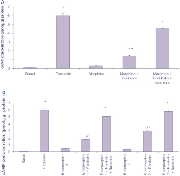
Methods: To test this hypothesis, we compared the basal expression of the mu (MOR), delta (DOR), and kappa (KOR) opioid receptors in the human U87 MG astrocytic cell line to SH-SY5Y neuronal and HL-60 immune cells using absolute quantitative real time RT-PCR (AQ-rt-RT-PCR). To demonstrate that IL-1β induced up-regulation of the MOR, DOR and KOR, U87 MG cells (2 x 10⁵ cells/well) were treated with IL-1β (20 ng/mL or 40 ng/mL), followed by co-treatment with interleukin-1 receptor antagonist protein (IL-1RAP) (400 ng/mL or 400 ng/mL). The above experiment was repeated in the cells desensitized with morphine, where U87 MG cells were pre-treated with 100 nM morphine. The functionality of the MOR in U87 MG cells was then demonstrated using morphine inhibition of forksolin-induced intracellular cAMP, as determined by radioimmunoassay.
Results: U87 MG cells treated with IL-1β for 12 h showed a significant up-regulation of MOR and KOR. DOR expression was also elevated, although not significantly. Treatment with IL-1β also showed a significant up-regulation of the MOR in U87 MG cells desensitized with morphine. Co-treatment with IL-1β and interleukin-1 receptor antagonist protein (IL-1RAP) resulted in a significant decrease in IL-1β-mediated MOR up-regulation.
Conclusion: Our results indicate that the pro-inflammatory cytokine, IL-1β, affects opiate-dependent pathways by up-regulating the expression of the MOR in both untreated and morphine-desensitized U87 MG.
Figures


Similar articles
-
Morphine and endomorphins differentially regulate micro-opioid receptor mRNA in SHSY-5Y human neuroblastoma cells.J Pharmacol Exp Ther. 2003 Aug;306(2):447-54. doi: 10.1124/jpet.103.048694. Epub 2003 May 16. J Pharmacol Exp Ther. 2003. PMID: 12754318
-
Dual regulation of mu opioid receptors in SK-N-SH neuroblastoma cells by morphine and interleukin-1β: evidence for opioid-immune crosstalk.J Neuroimmunol. 2010 Oct 8;227(1-2):26-34. doi: 10.1016/j.jneuroim.2010.06.007. Epub 2010 Jul 7. J Neuroimmunol. 2010. PMID: 20615556 Free PMC article.
-
Cholecystokinin receptor-1 mediates the inhibitory effects of exogenous cholecystokinin octapeptide on cellular morphine dependence.BMC Neurosci. 2012 Jun 8;13:63. doi: 10.1186/1471-2202-13-63. BMC Neurosci. 2012. PMID: 22682150 Free PMC article.
-
Morphine regulates gene expression of alpha- and beta-chemokines and their receptors on astroglial cells via the opioid mu receptor.J Immunol. 2002 Oct 1;169(7):3589-99. doi: 10.4049/jimmunol.169.7.3589. J Immunol. 2002. PMID: 12244149
-
Molecular targets of opiate drug abuse in neuroAIDS.Neurotox Res. 2005 Oct;8(1-2):63-80. doi: 10.1007/BF03033820. Neurotox Res. 2005. PMID: 16260386 Free PMC article. Review.
Cited by
-
Binge-Like Exposure to Ethanol Enhances Morphine's Anti-nociception in B6 Mice.Front Psychiatry. 2019 Jan 22;9:756. doi: 10.3389/fpsyt.2018.00756. eCollection 2018. Front Psychiatry. 2019. PMID: 30723430 Free PMC article.
-
Pomegranate-derived anthocyanin regulates MORs-cAMP/CREB-BDNF pathways in opioid-dependent models and improves cognitive impairments.J Ayurveda Integr Med. 2020 Oct-Dec;11(4):478-488. doi: 10.1016/j.jaim.2019.12.001. Epub 2020 May 16. J Ayurveda Integr Med. 2020. PMID: 32430240 Free PMC article.
-
Neuroimmune and Mu-Opioid Receptor Alterations in the Mesocorticolimbic System in a Sex-Dependent Inflammatory Pain-Induced Alcohol Relapse-Like Rat Model.Front Immunol. 2021 Sep 20;12:689453. doi: 10.3389/fimmu.2021.689453. eCollection 2021. Front Immunol. 2021. PMID: 34616393 Free PMC article.
-
General Anesthetics on Immune System Cytokines: A Narrative Review Article.Anesth Pain Med. 2020 Jul 5;10(4):e103033. doi: 10.5812/aapm.103033. eCollection 2020 Aug. Anesth Pain Med. 2020. PMID: 33134146 Free PMC article. Review.
References
-
- Sedqi M, Roy S, Mohanraj D, Ramakrishnan S, Loh HH. Activation of rat thymocytes selectively upregulates the expression of somatostatin receptor subtype-1. Biochem Mol Biol Int. 1996;38:103–112. - PubMed
Publication types
MeSH terms
Substances
Grants and funding
LinkOut - more resources
Full Text Sources
Research Materials
Miscellaneous
