Multicenter Study
. 2014 Jan;19(1):88-98.
doi: 10.1038/mp.2012.163. Epub 2012 Nov 20.

Clusterin regulates β-amyloid toxicity via Dickkopf-1-driven induction of the wnt-PCP-JNK pathway

Affiliations
Free PMC article
Multicenter Study

Clusterin regulates β-amyloid toxicity via Dickkopf-1-driven induction of the wnt-PCP-JNK pathway

R Killick et al. Mol Psychiatry. 2014 Jan.
Free PMC article

Abstract

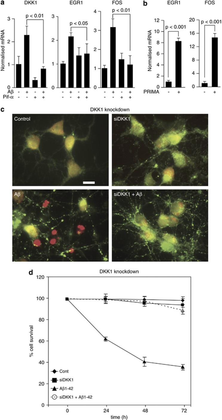
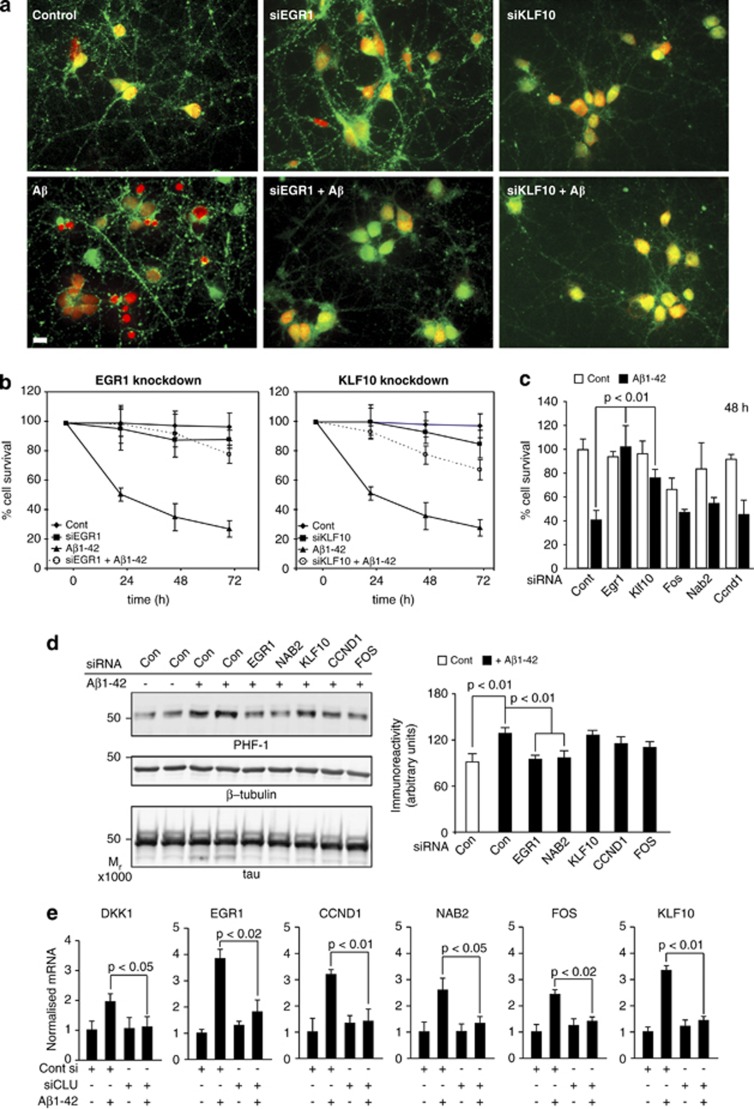
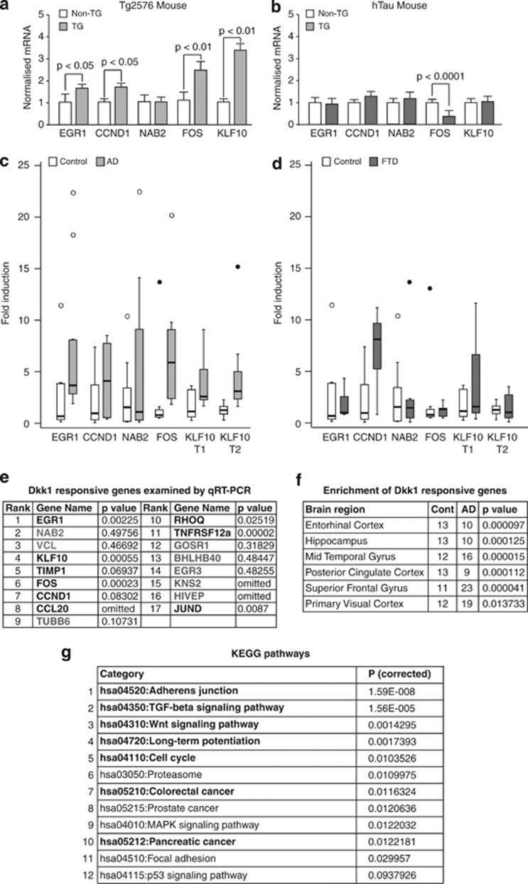
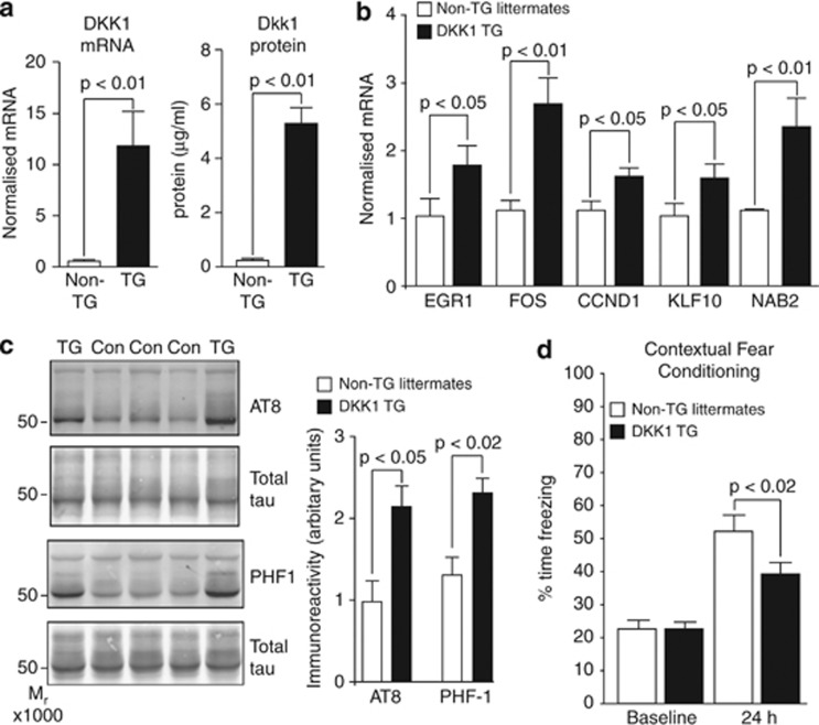
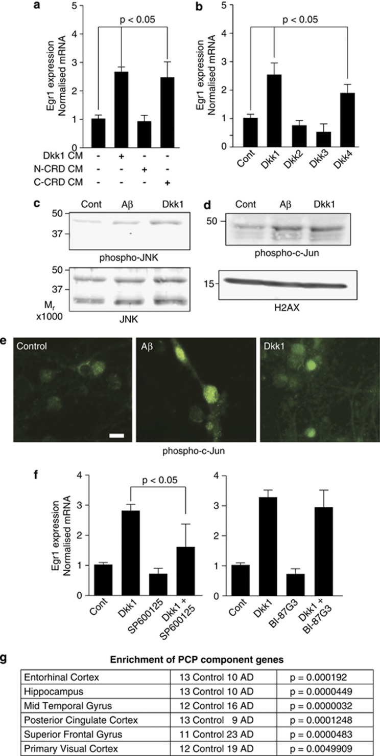
Although the mechanism of Aβ action in the pathogenesis of Alzheimer's disease (AD) has remained elusive, it is known to increase the expression of the antagonist of canonical wnt signalling, Dickkopf-1 (Dkk1), whereas the silencing of Dkk1 blocks Aβ neurotoxicity. We asked if clusterin, known to be regulated by wnt, is part of an Aβ/Dkk1 neurotoxic pathway. Knockdown of clusterin in primary neurons reduced Aβ toxicity and DKK1 upregulation and, conversely, Aβ increased intracellular clusterin and decreased clusterin protein secretion, resulting in the p53-dependent induction of DKK1. To further elucidate how the clusterin-dependent induction of Dkk1 by Aβ mediates neurotoxicity, we measured the effects of Aβ and Dkk1 protein on whole-genome expression in primary neurons, finding a common pathway suggestive of activation of wnt-planar cell polarity (PCP)-c-Jun N-terminal kinase (JNK) signalling leading to the induction of genes including EGR1 (early growth response-1), NAB2 (Ngfi-A-binding protein-2) and KLF10 (Krüppel-like factor-10) that, when individually silenced, protected against Aβ neurotoxicity and/or tau phosphorylation. Neuronal overexpression of Dkk1 in transgenic mice mimicked this Aβ-induced pathway and resulted in age-dependent increases in tau phosphorylation in hippocampus and cognitive impairment. Furthermore, we show that this Dkk1/wnt-PCP-JNK pathway is active in an Aβ-based mouse model of AD and in AD brain, but not in a tau-based mouse model or in frontotemporal dementia brain. Thus, we have identified a pathway whereby Aβ induces a clusterin/p53/Dkk1/wnt-PCP-JNK pathway, which drives the upregulation of several genes that mediate the development of AD-like neuropathologies, thereby providing new mechanistic insights into the action of Aβ in neurodegenerative diseases.


Figures

Figure 1
Figure 1
Aβ induction of Dickkopf-1 (Dkk1) is clusterin dependent. (a) Rat primary cortical neurons were treated with Pen1 small interfering RNA (siRNA) to CLU overnight and subsequently with 20 μM25-35 for 24 and 48 h and cell survival determined by the nuclear morphology assay. Cont, Control. (b) Neurons were treated as in (a), RNA collected after 3 h of Aβ treatment and qRT-PCR performed (detailed in Supplementary Methods). (c) Neurons were treated for 4 h with 20 μM25-35 and culture media and total cell lysates were collected and immunoblotted for clusterin. Clusterin in cell lysates was normalised to β-actin levels. (d) Neurons were treated with 20 μM25-35 for the times indicated and immunoblotted as in (c). Error bars in (c) and (d) show s.d. qRT-PCR, quantitative reverse transcription polymerase chain reaction.
Figure 2
Figure 2
Aβ-induced gene expression is dependent on p53 and is necessary and sufficient for neurotoxicity. (a) Rat cortical neurons were treated with 10 μM pifithrin-α for 18 h as indicated and subsequently with Aβ1-42(olig) (3 μM, 3 h). The expression levels of DKK1 (Dickkopf-1), EGR1 (early growth response-1) and FOS (FBJ murine osteosarcoma viral oncogene homologue) were determined by qRT-PCR. (b) Neurons were treated as in (b) using 10 μM PRIMA-1 and then with Aβ and qRT-PCR performed. (c) Rat neurons were treated o/n at 7 d.i.c. with control or Pen1 small interfering RNA (siRNA) to DKK1 (160 nM), and then with 3 μM1-42(olig) for 24 h and cytotoxicity assayed by the live/dead assay. Healthy cells are labelled green and dead cells are red. Scale bar=10 μM. (d) Neurons were treated as in (c) and cell survival determined at 24, 48 and 72 h by the nuclear morphology assay. d.i.c., days in culture; o/n, over night; qRT-PCR, quantitative reverse transcription polymerase chain reaction.
Figure 3
Figure 3
EGR1 (early growth response-1), KLF10 (Krüppel-like factor-10) and NAB2 (Ngfi-A-binding protein-2) mediate neurotoxicity and tau phosphorylation. (a) Neurons were treated o/n with Pen1 small interfering RNAs (siRNAs) to EGR1, FOS (FBJ murine osteosarcoma viral oncogene homologue), KLF10, NAB2, CCND1 (cyclin D1) and then with 3 μM1-42(olig) for 24 h and cytotoxicity assayed by the live/dead assay. Protective effects of siRNAs targeting EGR1 and KLF10 are shown. (b) Neurons were treated as in (a) and cell survival determined by the nuclear morphology assay up to 72 h. Significance values (not shown) for the effect of EGR1 and KLF10 siRNA on cell survival at each time point were ≤0.01. (c) Neurons were treated as in (a) and cell survival measured by lactate dehydrogenase (LDH) release. (d) Neurons were treated as in (a) and subsequently with 3 μM1-42(olig) for 4 h. Total lysates were collected and immunoblotted for phospho-tau using PHF-1. Immunoreactivity values for PHF-1 were normalised to total tau values; densitometric values are shown in the right. (e) Neurons were treated with Pen1-siCLU or control Pen1 siRNA and subsequently with 3 μM1-42(olig) for 3 h, RNA collected and qRT-PCR performed for DKK1 and the five Aβ/Dkk1 target genes. o/n, over night; qRT-PCR, quantitative reverse transcription polymerase chain reaction.
Figure 4
Figure 4
Aβ induced gene expression in amyloidopathy, but not tauopathy, brain. (a–d) Expression levels of the five common Aβ/Dickkopf-1 (Dkk1)-responsive genes were measured by qRT-PCR in total RNA from: cortex of (a) 12-month-old Tg2576 mice (n=9) and their non-TG littermate controls (n=7) and (b) 12-month-old hTau mice (n=5) and their non-TG littermate controls (n=5); and from hippocampi of (c) Alzheimer's disease (AD; n=10) and age-matched controls (n=9) and (d) frontotemporal dementia (FTD; n=9) and age-matched controls (n=9). TG, transgenic. Mouse data were normalised to HPRT and human data to GAPDH by the 2-ΔΔCT method. Significance was determined by one-way analysis of variance (ANOVA) and post hoc t-tests. Data are represented as normalised fold increases over control. Significance values of gene changes in human samples are given in table (e). Outlier values (between 1.5 and 3 times the interquartile range) and extreme values (>3 times the range) are shown as circles and filled circles, respectively. (e) Dkk1-responsive genes examined in AD hippocampus. Nonsignificant and omitted genes are shown in grey. (f) Human brain transcriptome data sets were mined with the top 50 most significant Dkk1-responsive genes. Significance of Dkk1 gene enrichment in AD in the six brain regions examined by Liang et al. are shown. Significance was determined by asymptotic globaltest (see Supplementary Information for full description). (g) KEGG (Kyoto Encyclopedia of Genes and Genomes) pathway analysis of human homologues of rat Dkk1-responsive genes. The pathway identifier, name and P-values after correcting for multiple testing by the method of Benjamini are shown. Pathways in bold have been associated with disease in AD brain expression data by Huang et al. qRT-PCR, quantitative reverse transcription polymerase chain reaction.
Figure 5
Figure 5
Transgenic overexpression of Dickkopf-1 (DKK1) induces ‘Aβ neurotoxicity pathway genes', tau phosphorylation and cognitive deficits. (a) DKK1 expression was determined by qRT-PCR (left bar graph) and enzyme-linked immunosorbent assay (ELISA; (right bar graph) in temporal cortex of neonatal DKK1 transgenic (TG) mice (n=6) and their non-transgenic (non-TG) littermates (n=5). (b) Neonatal expression of the five Aβ/Dkk1 genes determined by qRT-PCR. (c) Immunoblots of hippocampal lysates from 18- to 24-month-old DKK1 TG (n=7) and non-TG (n=7) mice using antibodies AT8 and PHF-1. Phosphoimmunoreactivity values were normalised to total tau values, within blot (bar charts, right). (d) The 14–16-month-old Dkk1 TG (n=17) and non-TG littermates (n=16) were subjected to contextual fear conditioning. Time spent freezing upon placement in the conditioning apparatus at baseline and at 24 h after training are shown. qRT-PCR, quantitative reverse transcription polymerase chain reaction.
Figure 6
Figure 6
The Aβ neurotoxicity pathway is the wnt–planar cell polarity (PCP) pathway. (a) Neurons were treated for 2 h with full-length, N-Terminal CRD or C-terminal CRD containing conditioned media (CM) and qRT-PCR performed. (b) Neurons were treated as in (c) with recombinant Dickkopf-1 (Dkk1), Dkk2, Dkk3 and Dkk4 proteins at 800 ng ml−1 and expression measured. (c) Neuronal lysates were immunoblotted for phospho-Thr183/Tyr185-SAPK/JNK and total SAPK/JNK following 3 h of treatments with 3 μM1-42(olig) or 800 ng ml−1 Dkk1. (d) Nuclear fractions were prepared from neurons treated as in (e) and immunoblotted for phospho-Serine63-c-Jun. Equal loading was determined using anti-H2A.X. (e) Neurons were treated as in (c), fixed and stained for phospho Ser63-c-Jun. Scale bar=10 μm. (f) Neurons were pre-treated with the c-Jun N-terminal kinase (JNK) inhibitors SP600125 and BI-87G3, each at 10 μM for 3 h, and then with 800 ng ml−1 recombinant Dkk1 for 2 h and qRT-PCR was performed. (g) Human brain transcriptomic data were mined with the wnt–PCP pathway component genes. Significance of differences in Alzheimer's disease (AD) in the six brain regions examined by Liang et al. are shown. CRD, cysteine rich domain; qRT-PCR, quantitative reverse transcription polymerase chain reaction; SAPK, stress activated protein kinase.

Similar articles

Cited by

References

    1. Glenner GG, Wong CW. Alzheimer's disease and Down's syndrome: sharing of a unique cerebrovascular amyloid fibril protein. Biochem Biophys Res Commun. 1984;122:1131–1135. - PubMed
    1. Brion JP, Flament-Durand J, Dustin P. Alzheimer's disease and tau proteins. Lancet. 1986;2:1098. - PubMed
    1. Hardy J, Allsop D. Amyloid deposition as the central event in the aetiology of Alzheimer's disease. Trends Pharmacol Sci. 1991;12:383–388. - PubMed
    1. Hardy J. The amyloid hypothesis for Alzheimer's disease: a critical reappraisal. J Neurochem. 2009;110:1129–1134. - PubMed
    1. Kremer A, Louis JV, Jaworski T, F VanLeuven. GSK3 and Alzheimer's disease: facts and fiction. Front Mol Neurosci. 2011;4:17. - PMC - PubMed

Publication types

MeSH terms