. 2013 Feb;21(2):445-55.
doi: 10.1038/mt.2012.234. Epub 2012 Nov 20.

Adipose-derived stromal cells overexpressing vascular endothelial growth factor accelerate mouse excisional wound healing

Affiliations

Adipose-derived stromal cells overexpressing vascular endothelial growth factor accelerate mouse excisional wound healing

Allison Nauta et al. Mol Ther. 2013 Feb.

Abstract

Angiogenesis is essential to wound repair, and vascular endothelial growth factor (VEGF) is a potent factor to stimulate angiogenesis. Here, we examine the potential of VEGF-overexpressing adipose-derived stromal cells (ASCs) for accelerating wound healing using nonviral, biodegradable polymeric vectors. Mouse ASCs were transfected with DNA plasmid encoding VEGF or green fluorescent protein (GFP) using biodegradable poly (β-amino) esters (PBAE). Cells transfected using Lipofectamine 2000, a commercially available transfection reagent, were included as controls. ASCs transfected using PBAEs showed enhanced transfection efficiency and 12-15-fold higher VEGF production compared with cells transfected using Lipofectamine 2000 (*P < 0.05). When transplanted into a mouse wild-type excisional wound model, VEGF-overexpressing ASCs led to significantly accelerated wound healing, with full wound closure observed at 8 days compared to 10-12 days in groups treated with ASCs alone or saline control (*P < 0.05). Histology and polarized microscopy showed increased collagen deposition and more mature collagen fibers in the dermis of wound beds treated using PBAE/VEGF-modified ASCs than ASCs alone. Our results demonstrate the efficacy of using nonviral-engineered ASCs to accelerate wound healing, which may provide an alternative therapy for treating many diseases in which wound healing is impaired.


Figures

Figure 1
Figure 1
Optimizing gene delivery to ASCs using biodegradable polymeric vectors. (a) Transfection efficiency was visualized using fluorescence microscopy (left) and quantified with FACS analysis for GFP production (middle and right). FACS analysis generally supported a 4 µg optimal treatment dose, with efficiencies ranging from 6 to 10%. A one-way ANOVA was used to test for differences among groups, showing statistically significant differences across the eight groups, F (7, 16) = 101.6, *P < 0.05. Tukey post-hoc comparisons showed that cells treated with the 4 µg treatment dose consistently demonstrated twofold higher transfection efficiency than cells transfected with Lipofectamine 2000. (Mean difference = -3.5, 99.9% CI of difference = -5.356 to -1.644, ***P < 0.001). All experiments were performed in triplicates and were repeated nine times to confirm findings. (b) VEGF ELISA demonstrated increased VEGF protein production from PBAE/VEGF transfected cells on day 2 post-transfection. One-way ANOVA showed statistically significant differences overall between the eight groups, F (7, 16) = 31.23, *P < 0.05. At the optimal dose (4 µg), PBAE transfection produced 12–15 times higher VEGF protein than Lipofectamine 2000, as determined by Tukey post-hoc comparison (mean difference = -6,807, 99.9% CI of difference = -10,656 to 2,957, ***P < 0.001). With higher DNA load, VEGF concentration decreased, as demonstrated by 4 versus 6 µg loading doses (mean difference = 3,143, 99% CI of difference = 129.7 to 6,156, **P < 0.01). (c) Cell viability was slightly lower with transfection with any reagent compared to the untreated group (**P < 0.01, as applied to every treatment group compared with untreated), but viability after transfection with PBAE at any dose was not statistically significantly different from viability after transfection with Lipofectamine 2000, as evaluated by one-way ANOVA and Tukey post-hoc comparison. All values are expressed as mean ± SEM (n = 3). ANOVA, analysis of variance; CI, confidence interval; FACS, fluorescence-activated cell sorting; GFP, green fluorescent protein; PBAE, poly (β-amino) esters; VEGF, vascular endothelial growth factor.
Figure 2
Figure 2
In vitro matrigel tubulogenesis assay. HUVECs treated with conditioned media from PBAE/VEGF-transfected ASCs showed significantly increased tubulogenesis compared to cells treated with conditioned media collected from untreated ASCs. Differences were statistically significant, as evaluated by one-way ANOVA and Tukey post-hoc test. ANOVA: F (2, 20) = 16.70; ***P < 0.001; Tukey (untreated versus PBAE): mean difference: -4,226, ***P < 0.001, 99.9% CI of difference: -7,361 to -1,090; Tukey (Lipo versus PBAE): mean difference: -2,321,*P < 0.05, 95% CI of difference: -4,171 to -471.0. Values are expressed as mean ± SEM (n = 10 high-powered fields per well, three wells per group). ANOVA, analysis of variance; ASC, adipose-derived stromal cell; CI, confidence interval; HUVEC, human umbilical vein endothelial cell; PBAE, poly (β-amino) esters; VEGF, vascular endothelial growth factor.
Figure 3
Figure 3
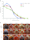
Wound healing in a mouse excisional wound model. (a) Percent of open wound was evaluated every 2 days post-wounding. PBAE/VEGF-modified ASCs produced significantly accelerated wound healing compared to groups treated with ASCs or PBS control. Values expressed as mean ± SEM (n = 20 wounds total per group, 10 mice per group). Statistics were performed on each day using one-way ANOVA and showed statistically significant differences between groups from days 2–10. For days 2, 8, and 10, all three combinations of groups (PBS and ASC, PBS and PBAE, and ASC and PBAE) were statistically significant with *P < 0.05 or better, as evaluated by Tukey post-hoc test. (b) Representative images show accelerated wounds closure in group treated with PBAE/VEGF-transfected ASCs, with full epithelialization observed by day 8. ANOVA, analysis of variance; ASC, adipose-derived stromal cell; PBAE, poly (β-amino) esters; PBS, phosphate-buffered saline; VEGF, vascular endothelial growth factor.
Figure 4
Figure 4
Evaluation of inflammation. (a) May-Grünwald Giemsa quantification of leukocytes from wounds harvested on day 2 show increased leukocyte recruitment in the dermis on either side of wounds in the PBAE/VEGF-transfected ASC group compared to both the untreated ASC and PBS control groups (**P < 0.05). (n = 3 wounds per group, six regions of interest per group). Differences between ASC- and PBS-treated controls were not statistically significant, as evaluated by one-way ANOVA and Tukey post-hoc test. (b) From left to right, representative images showing hematoxylin and eosin stains (×20) of the wound edges showing dermal cellularity, May-Grünwald Giemsa staining for leukocytes and the wound borders (×20), and higher magnification of May-Grünwald Giemsa-stained tissue, demonstrating marked leukocytosis in tissue treated with PBAE/VEGF-transfected ASCs. Yellow arrow points to leukocytes. Bars: 20 µm. (c) Immunofluorescence of F4/80 stained tissue sections on day 2 show a high density of macrophages at the periphery of wounds treated with PBAE/VEGF-transfected ASCs. Red arrows points to macrophages. Bars: 20 µm. ANOVA, analysis of variance; ASC, adipose-derived stromal cell; DAPI, 4′,6-diamidino-2-phenylindole; PBAE, poly (β-amino) esters; PBS, phosphate-buffered saline; VEGF, vascular endothelial growth factor.
Figure 5
Figure 5
VEGF ELISAs of skin tissue lysates. (a) Human VEGF ELISA of skin tissue lysates from day 1 show high levels of human VEGF protein production in wounds treated with PBAE/VEGF-transfected ASCs, versus negligible levels in ASC- and PBS-treated controls (***P < 0.001 for comparisons of PBAE/VEGF versus ASC and PBAE/VEGF versus PBS; ANOVA: F (2, 15) = 198.8. Tukey mean difference: 818.8 and 827.0; 99.9% CI of difference = 600.8 to 1,037 and 609.0 to 1,045, respectively). (b) Mouse VEGF ELISA showed elevated levels of mouse VEGF in tissue treated with PBAE/VEGF-transfected ASCs compared to ASC- and PBS-treated controls (**P < 0.01 and ***P < 0.001, respectively. ANOVA: F (2, 15) = 26.43. Tukey mean difference: 146 and 275, 99% CI of difference = 16.31 to 275.7 and 145.9 to 405.3). Differences between ASC- and PBS-treated controls were not statistically significant. For each group, n = 2 wounds evaluated. Each sample was run in triplicate. All values are expressed as mean ± SEM. Statistical analysis was evaluated using one-way ANOVA and Tukey post-hoc test. ANOVA, analysis of variance; ASC, adipose-derived stromal cell; CI, confidence interval; PBAE, poly (β-amino) esters; PBS, phosphate-buffered saline; VEGF, vascular endothelial growth factor.
Figure 6
Figure 6
Evaluation of blood vessel formation. (a) CD31 labeling of endothelial cells (red) and fluorescence microscopy at ×20 shows increased staining on day 14 in the wound and surrounding tissue in the PBAE/VEGF ASC-treated group compared with ASC and PBS controls (**P < 0.01 and *P < 0.05, respectively; n = 3 wounds evaluated per group; ANOVA: F (2, 8) = 10.72; Tukey mean difference: -10.40 and 10.69; 95% CI of difference: -17.4 to -3.40 and 2.86 to 18.5, respectively). All values are expressed as mean ± SEM. Quantification (left) and representative images (right). White arrows point to endothelial cells. (b) Hematoxylin and eosin (H&E) and Verhoeff-Van Gieson (V-VG) stains were used to evaluate quality of blood vessels. From left to right, H&E and V-VG stains of dermal scar show blood vessels forming a continuous networks parallel to the skins' surface. H&E and V-VG of skin flanking dermal scar demonstrates mature blood vessels coursing parallel to the skin's surface. Black arrows point to blood vessels. Bars: 20 µm. ANOVA, analysis of variance; ASC, adipose-derived stromal cell; CI, confidence interval; DAPI, 4′,6-diamidino-2-phenylindole; PBAE, poly (β-amino) esters; PBS, phosphate-buffered saline; VEGF, vascular endothelial growth factor.
Figure 7
Figure 7
Cell survival post-transplantation in vivo. (a) Bioluminescence imaging showed comparable cell engraftment 24 hours post-transplantation (right), with prolonged cell survival detected in PBAE/VEGF-transfected ASCs between days 6 and 10 (*P < 0.05; left). Statistical analysis was performed using Student's t-test between the two ASC groups; PBS injection was included as negative control and is shown in this figure only to illustrate background luminescence. Values expressed as mean ± SEM (n = 6). (b) Representative images from bioluminescence imaging studies on day 6 post-wounding showed higher level of cell viability in groups treated with PBAE/VEGF ASCs than ASCs alone. (c) Immunofluorescence shows increased cellularity in groups treated with PBAE/VEGF/ASCs on day 4 post-wounding. Transplanted cells (labeled green: anti-luciferase) frequently colocalized in proximity to blood vessels (labeled red: anti-CD31). ASC, adipose-derived stromal cell; DAPI, 4′,6-diamidino-2-phenylindole; PBAE, poly (β-amino) esters; PBS, phosphate-buffered saline; VEGF, vascular endothelial growth factor.
Figure 8
Figure 8
Histology of wound beds day 14 post-treatment. (a) Hematoxylin and eosin and Masson's trichrome staining shows increased cellularity and collagen deposition in the dermis treated with both ASC-transplanted groups than PBS-treated control. (b) PBAE/VEGF/ASC-treated group shows most abundant mature collagen fiber (red-orange bifringent, by Picrosirius red staining), whereas PBS-treated group showed highest level of immature collagen fiber (green-yellow bifringent, by polarization microscopy). ASC, adipose-derived stromal cell; PBAE, poly (β-amino) esters; VEGF, vascular endothelial growth factor.

Similar articles

Cited by

References

    1. Gurtner GC, Werner S, Barrandon Y., and, Longaker MT. Wound repair and regeneration. Nature. 2008;453:314–321. - PubMed
    1. Sen CK, Gordillo GM, Roy S, Kirsner R, Lambert L, Hunt TK.et al. (2009Human skin wounds: a major and snowballing threat to public health and the economy Wound Repair Regen 17763–771. - PMC - PubMed
    1. Galiano RD, Tepper OM, Pelo CR, Bhatt KA, Callaghan M, Bastidas N.et al. (2004Topical vascular endothelial growth factor accelerates diabetic wound healing through increased angiogenesis and by mobilizing and recruiting bone marrow-derived cells Am J Pathol 1641935–1947. - PMC - PubMed
    1. Galeano M, Deodato B, Altavilla D, Cucinotta D, Arsic N, Marini H.et al. (2003Adeno-associated viral vector-mediated human vascular endothelial growth factor gene transfer stimulates angiogenesis and wound healing in the genetically diabetic mouse Diabetologia 46546–555. - PubMed
    1. Kirchner LM, Meerbaum SO, Gruber BS, Knoll AK, Bulgrin J, Taylor RA.et al. (2003Effects of vascular endothelial growth factor on wound closure rates in the genetically diabetic mouse model Wound Repair Regen 11127–131. - PubMed

Publication types

MeSH terms

LinkOut - more resources